
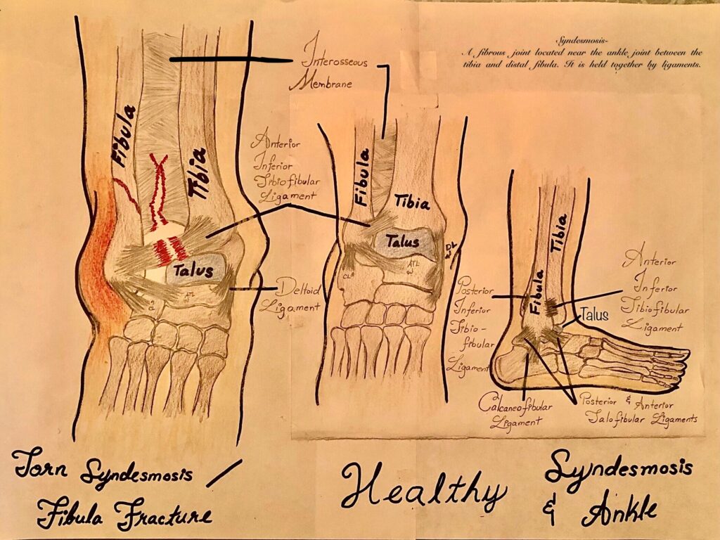
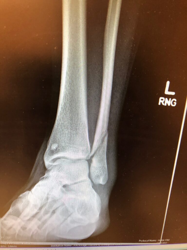
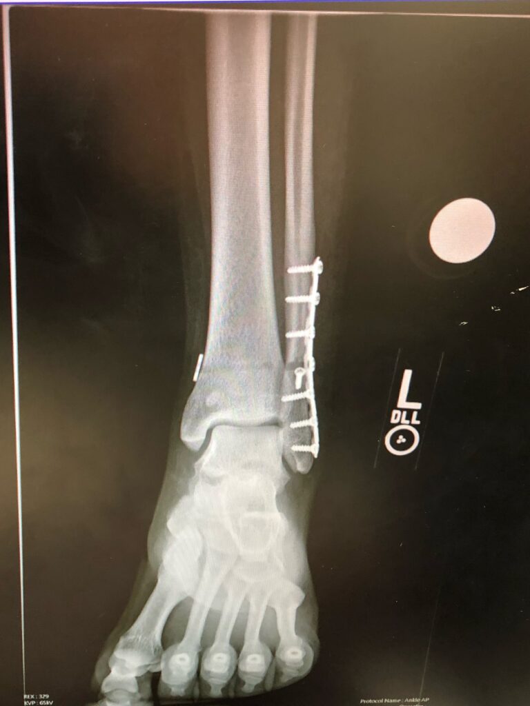
For my STEAM project, I chose to do syndesmosis injury that results from fibular fractures and the use of open reduction and internal fixation (ORIF) to help achieve optimal bone repair. This primarily relates to the objective “know the stages of bone development and repair”. For my media, I’m presenting X-rays to provide a visual of ORIF, specifically internal fixation, and an illustration of the fibula, tibia, and syndesmosis joint (normal and healthy compared to torn and fractured).
The syndesmosis is a fibrous joint more formally known as the distal tibiofibular syndesmosis. As its name implies, this joint is located near the ankle, between the tibia and distal fibula. The four ligaments holding this joint in place are the anterior inferior tibiofibular ligament, the posterior inferior tibiofibular ligament, the interosseous ligament, and the transverse tibiofibular ligament. A syndesmotic injury is a condition in which there has been damage to these ligaments connecting and stabilizing the ankle, tibia, and fibula. Most often, syndesmotic injuries result from fractures of the tibia or fibula. It occurs in the event of a twisting or rotational injury. If the injury remains uncorrected, it can lead to chronic instability, significant morbidity, and ultimately lead to degenerative arthritis. To prevent the increasing risk of potentially developing these conditions, the injury and fracture can be healed with the use of ORIF.
ORIF is an acronym for open reduction and internal fixation. It is a form of surgery used to stabilize and heal broken bones. During open reduction, orthopedic surgeons will reposition the pieces of the bone and restore them to proper alignment. On the other hand, internal fixation has to do with physically reconnecting the bones and may involve special screws, plates, rods, wires, or nails that the surgeon inserts to fix bones in the proper position. This prevents abnormal healing and usually takes place while the patient is under general anesthesia.

Fixed
After reading and seeing the completed project, the process to repair a syndesmotic injury is very apparent. I have learned that the lower leg is composed of the tibia, fibula and their supporting ligaments. The bones and ligaments provide a strong structure, so injury to this area can be detrimental. Injury of these ligaments can be known as a syndesmotic injury and can be fixed by open reduction or internal fixation: open reduction being when the bones are realigned and internal fixation is when the bones require screws, bolts, etc, for structural support. The process of “fixing” the injury will begin after phagocytes clear the injury of debris and bacteria. Once this has occurred, fibroblasts will repair tissue and osteocytes will repair bone. The X-ray images Lynciemay used show a clear difference between a bone in good condition and a bone after being repaired. The additional illustration also helped localize what bones and ligaments are a part of a syndesmotic injury.