
I will be researching how tattoo ink is dispersed into the skin. The course objective it will relate to is the functions of the integumentary system. All of the main parts of the integumentary system that are mentioned are labeled in the visual art.
The integumentary system is the biggest organ in the body that serves to create a safety barrier between the world around us and our internal body. The integumentary system includes the epidermis, dermis, hypodermis, associated glands, hair, and nails (Kim & Dao, 2023). The picture of the skin in this project doesn’t show the hair follicles because tattoos usually don’t affect them. The visual for this project will display all the parts of the skin; the epidermis, the dermis, and the hypodermis. There are two main layers of tissue from the integumentary system that makeup the skin.
The first layer is known as the epidermis and the second layer is known as the dermis. The epidermis is the part of our skin that is visible. It is represented by the color peach in the visual art piece. It is made up of stratified keratinised squamous epithelium which can be explained by breaking down each phrase of the tissue. Epithelium is a sheet of cells that covers body surfaces and cavities. It appears in two forms which are covering/lining epithelium and glandular epithelia. Covering and lining epithelia are on external and internal surfaces such as the skin. Glandular epithelium are in secretory tissues such as saliva glands. The covering and lining epithelia function to protect our body from harm which is exactly what skin does. Epithelial tissue is avascular but innervated. Avascular means there are no blood vessels in the epidermis tissue. That is why when you get small cuts such as a paper cut, there isn’t any blood. Innervated means there are still nerves attached to the tissue. The nerves in the visual art are aqua and extend from the hypodermis into the epidermis. The blood vessels are contained in the layer beneath; the dermis. All epithelial tissues have two names; simple epithelium and stratified epithelium. All this refers to is the number of cell layers thick the tissue is. Simple means one layer thick while stratified means it is multiple layers thick. Keratinised means that the cells are hardened and found in the skin. Finally the term squamous refers to the shape of the cells. Squamous means they are flattened and scale-like. So to summarize, the epidermis is made up of epithelial cells that are multiple layers thick, located in the skin, and are flat cells.
The layer beneath, the dermis, is composed of two different layers of cells; the reticular layer and the papillary layer. The papillary is pink in the art piece. The papillary contains the blood and nerves that help maintain the epidermis. Capillaries are the smallest blood vessels we have. The red and blue strands in the art piece represent the capillaries. The reticular layer is made up of collagen and elastic fibers that are woven together to help the skin be able to stretch and contract. The reticular is white because there are so many things in it so I kept it uncolored. The hypodermis is a third layer. It is not part of the skin, but the skin rests on this layer, which attaches it to underlying bone and muscle. It primarily consists of fat cells and connective tissue, serving as a cushion and insulation for the body. In the art piece the fat cells are depicted with yellowish ovals and the red orange is the connective tissue.
The ink is injected between the papillary and reticular layer. The ink is depicted in the art piece in multiple places under the papillary layer including at the end of the needle and it is purple. The reason it is injected into the reticular layer is to bypass the outer layer of the skin, the epidermis. The epidermis constantly renews everyday so the needle must inject to a spot that won’t lose the ink right away. The papillary layer contains capillaries that supply nutrients to the epidermis and nerve endings essential for sensation. The capillaries extend from the hypodermis to the papillary layer in the art piece. Placing ink in the reticular layer ensures longevity and visibility of the tattoo, as it is deep enough to resist the constant renewal of the epidermis.
Resources
- Grant, C. A.; Twigg, P. C.; Baker, R.; Tobin, D. J. Beilstein J. Nanotechnol. 2015, 6, 1183–1191 https://doi.org/10.3762/bjnano.6.120
- McLafferty, E., Hendry, C., & Farley, A. (2012). The integumentary system: Anatomy, physiology and function of skin. Nursing Standard (through 2013), 27(3), 35-42. Retrieved from http://uaf.idm.oclc.org/login?url=https://www.proquest.com/scholarly-journals/integumentary-system-anatomy-physiology-function/docview/1081981576/se-2
- Kim, J. Y., & Dao, H. (2023, May 1). Physiology, integument – statpearls – NCBI bookshelf. National Library Of Medicine. https://www.ncbi.nlm.nih.gov/books/NBK554386/
- Hewings-Martin, Y. (2017b, July 14). Tattoos: Does ink travel through your body?. Medical News Today. https://www.medicalnewstoday.com/articles/318388#Pigments-are-lodged-in-the-dermis
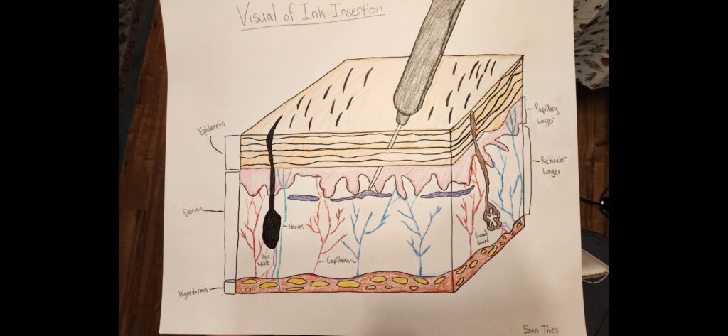
Sean’s post was about how ink for tattoo’s stay within people’s skin. The answer to this is that the ink is injected into the dermis. The dermis is the second layer of skin and is below the epidermis. As the epidermis is made up of epithelial tissue and would regenerate quickly causing the ink to disappear. This makes it a poor choice for the retention of the ink. Instead of injecting into the epidermis we inject the ink into the dermis. The dermis does not die and regeneration as fast as the dermis does and therefore retains the ink better. Sean’s picture explains this well by showing how the needle entirely bypasses the epidermis.