
The objectives related to my steam project is to compare and contrast the peripheral and central nervous system and compare and contrast the various nervous tissues and cells. The brain is part of the central nervous system and is located in the cranial cavity. The cranial cavity is made by a hard nonflexible skull and has little room to accommodate a growing mass. Tumors that grow in the brain, whether benign or malignant, or an accumulation of any substance within the skull is never good because of this limited space. Once there is no more room for the growth to be accommodated, intracranial pressure starts to build up. Intracranial pressure building up can cause extreme pain, and optic nerve damage. More seriously it can also crush brain tissue, cut off blood vessels, and even push part of the brain through the hole in the base of the skull called the foramen magnum where the spinal cord connects to the brain which causes irreversible brain stem damage. These complications can cause brain damage, stroke, paralysis, coma, and death.
Ependymoma is a type of glioma(brain tumor). They are a rare tumor that is most common in children under ten. Ependymoma accounts for eight to ten percent of brain tumors in that age group as opposed to 1.7% of brain tumors in all ages. Ependymomas occur in the ependymal cells that make up the ependyma that lines the central canal of the spinal cord or in the ependymal cells of the ependyma of the cerebral ventricle. In children, ependymomas most often occur near the cerebellum in the ependyma around the 4th ventricle, either in the 4th ventricle or in the cerebral aqueduct. If an ependymoma in this area grows to a sufficient size it can block the circulation of cerebrospinal fluid. With the 4th ventricle area blocked or severely restricted cerebrospinal fluid can not continue its circulation and will begin to build up in the 3rd ventricle and the lateral ventricles also known as the 2nd and 1st ventricles. This buildup of cerebrospinal fluid in the cerebral ventricle is known as intraventricular obstructive hydrocephalus, also known as non-communicating hydrocephalus. Hydrocephalus means, in Latin, water on the brain. This aptly describes the condition of cerebrospinal fluid building up in the brain. The cerebrospinal fluid slowly accumulates and builds up intracranial pressure. Which as shown earlier in the essay is bad.
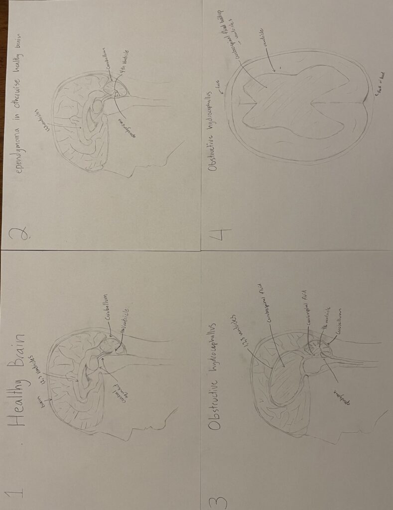
The first sketch shows an MRI of a healthy tumor and hydrocephalus free brain from the sagittal view. The brain, cerebral ventricles, cerebral aqueduct, and cerebellum are labeled. The second sketch shows an Mri of an otherwise healthy brain with the formation of an ependymoma. The ventricles, the ependymoma, and the cerebellum are labeled. The third sketch shows a sagittal brain MRI of a brain with obstructive hydrocephalus. The lateral, 3rd, 4th ventricle, and cerebral aqueduct are all enlarged with the buildup of the cerebrospinal fluid in the brain from the ependymoma caused blockage. The ventricles, the aqueduct, and the cerebellum are labeled. The fourth sketch shows a CT scan from the axial view of a brain with obstructive hydrocephalus. The ventricles are labeled.
Ependymomas and hydrocephalus helps with a deeper understanding of the objectives by showing how hydrocephalus affects and harms the CNS. Ependymomas go into understanding nervous tissue by talking about the tumor’s tissue type, and help with understanding the CNS and how the tumor blockage affects the ventricles and the circulation of cerebrospinal fluid.
Citations
Andreas, A. Obstructive Hydrocephalus – an overview | ScienceDirect Topics, 2021. Www.sciencedirect.com. https://www.sciencedirect.com/topics/medicine-and-dentistry/obstructive-hydrocephalus
Ependymoma: Danger in the fourth ventricle. (2011, August 25). Pathology Student. https://www.pathologystudent.com/ependymoma-danger-in-the-fourth-ventricle/
“Gliomas.” Gliomas | Johns Hopkins Medicine, John Hopkins Medical, 11 Feb. 2022, https://www.hopkinsmedicine.org/health/conditions-and-diseases/gliomas
Hydrocephalus due to congenital stenosis of aqueduct of sylvius – About the Disease – Genetic and Rare Diseases Information Center. (n.d.). Rarediseases.info.nih.gov. Retrieved November 23, 2022, from https://rarediseases.info.nih.gov/diseases/434/hydrocephalus-due-to-congenital-stenosis-of-aqueduct-of-sylvius
Hydrocephalus. (n.d.). Www.meddean.luc.edu. Retrieved November 23, 2022, from http://www.meddean.luc.edu/lumen/MedEd/Radio/curriculum/Neurology/Hydrocephalus_2013.htm
Latham, K. (2021, April 21). Ependymal Cell – The Definitive Guide. Biology Dictionary. https://biologydictionary.net/ependymal-cells/
Pressman, P. Overview of Elevated Intracranial Pressure. (n.d.). Verywell Health. Retrieved November 23, 2022, from https://www.verywellhealth.com/elevated-intracranial-pressure-2488707#:~:text=If%20pressure%20builds%20above%20the%20membrane%2C%20brain%20tissue
Sa, M. (2016). The Spinal Ependymal Layer in Health and Disease. Veterinary Pathology, 53(4). https://doi.org/10.1177/0300985815618438
Sharma, A., & Graber, J. J. (2021). Overview of prognostic factors in adult gliomas. Annals of Palliative Medicine, 10(1), 863–874. https://doi.org/10.21037/apm-20-640Zamora, E. A., & Fahad Alkherayf. (2019, November 16). Ependymoma. Nih.gov; StatPearls Publishing. https://www.ncbi.nlm.nih.gov/books/NBK538244/
The focus of Owen Averett’s STEAM project was understanding ependymoma and how it interacts with the peripheral and central nervous system and the various nervous tissues and cells involved. Ependymoma is a type of glioma or brain tumor that grows from the ependymal cells that line the central canal of the spinal cord or in the ependyma of the cerebral ventricle. Found most often in children under the age of ten. This cancer grows near the cerebellum in the ependyma around the 4th ventricle, or cerebral aqueduct. This disease accounts for eight to ten percent of all gliomas in children under ten as opposed to seventeen percent in all age groups. With malignant growth in these regions, we can see restriction of cerebrospinal fluid movement; the decrease in this movement leads to increased intracranial pressure in the third, second, and first ventricles respectively. This buildup is referred to as intraventricular obstructive hydrocephalus or non-communicating hydrocephalus as it can lead to cognitive or physical development. This cognitive or physical development impairment is due to an increase in intracranial pressure stemming from the inflexibility of the cranial vault. In moderate cases this intracranial pressure will likely lead to, pain and optic nerve damage. However, in more serious cases or cases left untreated, ischemia of brain tissue, reduction or obstruction of the local to regional blood vessels, and eventually herniation of the brain stem through the foramen magnum and consequentially irreversible damage to the brain stem. This damage will lead to stroke, paralysis, coma, or death of the patient.
Thank you for writing about this, it’s an interesting topic.