


The objective that I chose to focus on is “know the location of bones listed”. This one sounds easy enough, but the human skull has twenty two bones alone. At the beginning of this unit, I really only knew about the mandible, the frontal bone, the temporal bone and the orbital canal. I never really thought of the skull having that many bits and pieces. I also didn’t think that I would enjoy learning about the skeleton as much as I did, specifically the skull.
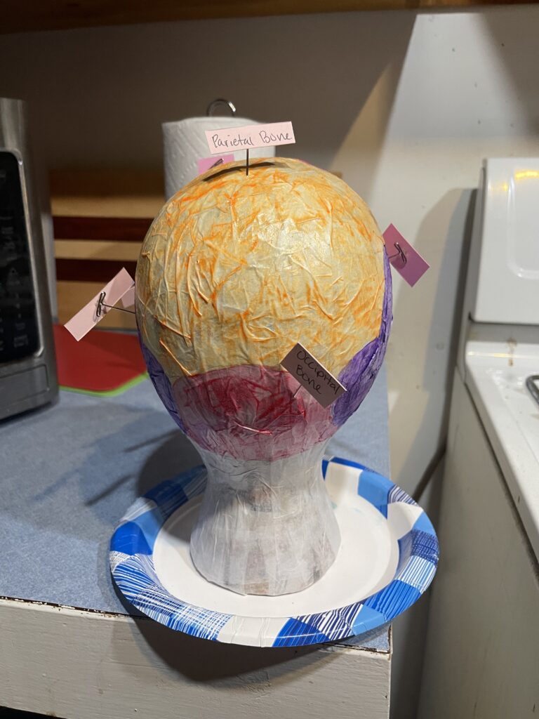
The skull is a part of the axial skeleton which forms the vertical axis of the body. This also includes the vertebral column and the thoracic cage. The skull is more specifically known as the cranium. It has two main parts; the braincase and the facial bones. The facial bones are located at the anterior of the skull, the brian case is shown by just about every other view. The main function of the brain case is to protect and contain the brain. The cranial cavity, which is where the brain is located, is bounded by the calvaria (the skullcap) and the lateral and posterior sides of the skull. The brain case is made of the frontal bone, parietal bones, occipital bone, and the sphenoid bone. The sphenoid bone is a part of the central skull and it joins with almost every other bone in the skull.
The anterior structure of the skull mostly includes the glabella, nasal cavity, orbit, mandible, maxilla, and the zygomatic bone. The nasal cavity is split into two halves via the nasal septum. The interior of the nasal septum consists of the perpendicular plate of the ethmoid bone and below that is the vomer bone. The rest of the nasal cavity is formed by three plates. From the front view there is a larger plate, the inferior nasal concha, which is an independent bone in the skull and then the middle nasal concha, which is a part of the ethmoid bone. The third plate you can’t see, it’s much smaller and located above the middle concha. This one is known as superior nasal concha. Above all of this is the nasal bone.
The orbit is the bony socket where the eyeball and surrounding muscles are located. The upper portion of the anterior orbit is the supraorbital margin. Closer to the midpoint of the supraorbital margin is a small opening called the supraorbital foramen. Just below the orbit is the infraorbital foramen. This is where the point of emergence for a sensory nerve for the face is located. Also included in the orbit is the ethmoid bone, part of the sphenoid bone, the palatine bone and part of the lacrimal bone.
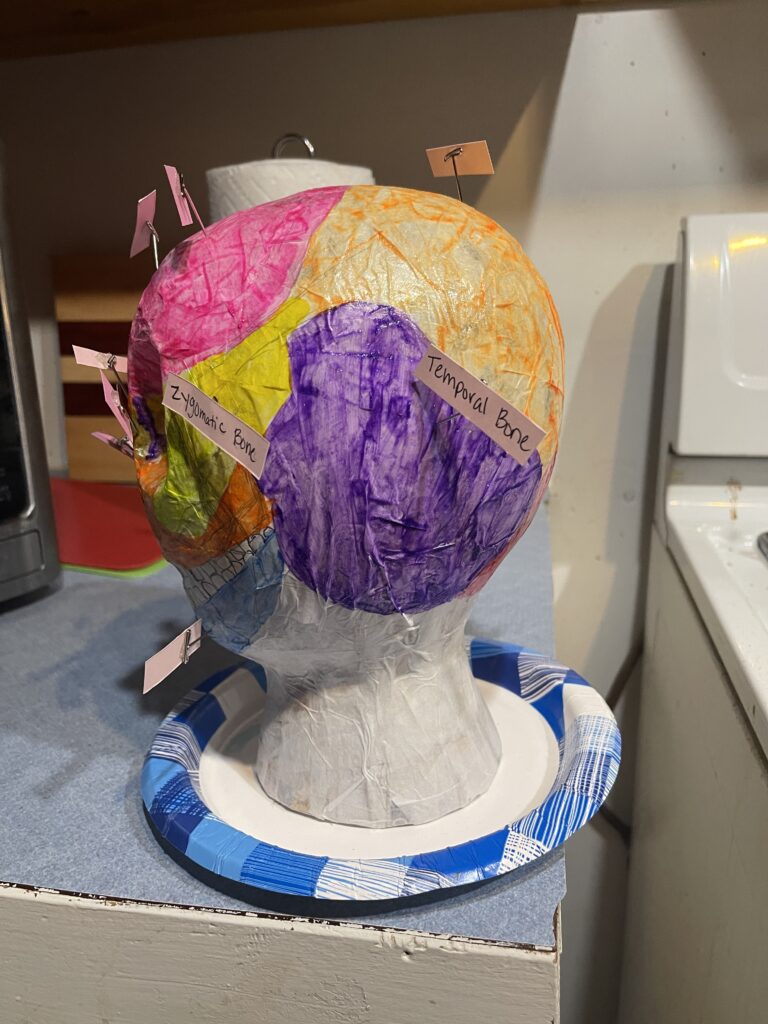
The lateral view of the skull is where you can largely see the brain case above and the upper and lower jaws with their teeth below. The bone that separates these two areas is called the zygomatic arch. It spans from the area of the cheek ti just above the ear canal. It is connected by two processes: the temporal process of the zygomatic bone and the zygomatic process of the temporal bone. The bones of the brain case largely consist of the parietal bone, the temporal bone, the frontal bone and glabella, and the occipital bone. The parietal, occipital, temporal, and zygomatic bones can all be seen from the posterior view.
As you can see the human skull is vastly more complicated than it may appear. It includes large bones and many smaller processes as well.