Compare and contrast the organelles of compact bone
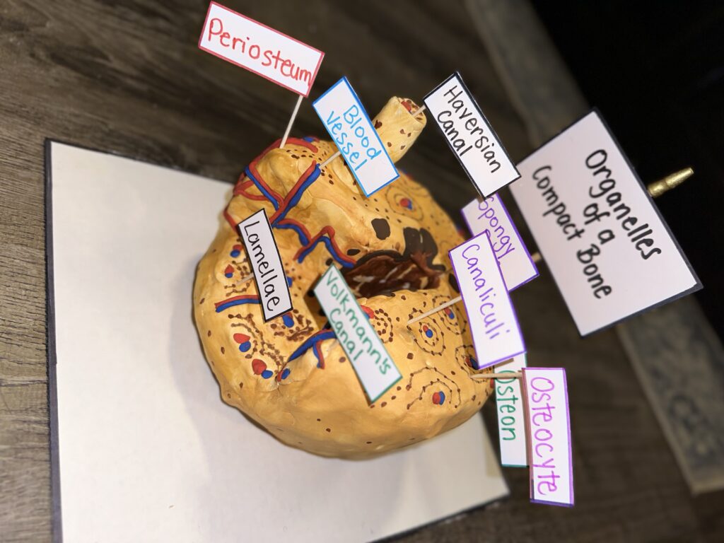
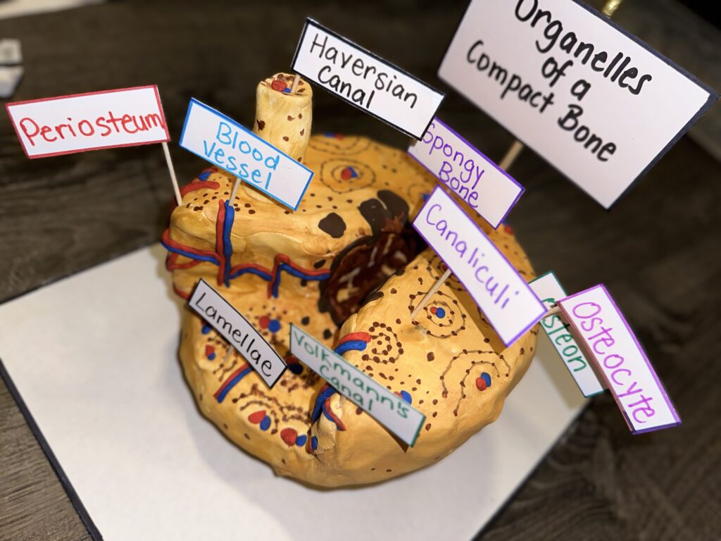
The STEAM project represents 3D art and craft techniques combined with paintings and drawings of the organelles of the Compact bone tissue also called the Cortical bone. To further elaborate, the organelles are labeled for easy identification by viewers of this project. The objective is to Compare and contrast the organelles and their function. Osteomyelitis is a disease that is associated with Cortical bone. Bone is a complicated organ that supports various physiological activities and serves multiple functions like body support, movement facilitation, organ protection, mineral and fat storage, and hematopoiesis. Bone loss resulting from diseases, trauma, tumor, or aging impacts the life quality and even the survival of an individual. It was reported that bone grafting represented more than 2 million worldwide each year, making bone the second most common transplant tissue (Chang, 2022).
First, the art displayed above points out the organelles associated with compact bone tissue. The hard outer layer of bones, known as compact bone or cortical bone, surrounds the bone marrow. It offers protection and strength to bones and is made up of units called osteons or Haversian systems. Osteons are cylindrical structures that consist of a mineral matrix and living osteocytes connected by canaliculi, which transport blood. They are aligned parallel to the bone’s long axis. Each osteon is made up of layers of compact matrix called lamellae, which surround a central canal called the Haversian canal. The Haversian canal contains the bone’s blood vessels and nerve fibers. Osteons in compact bone tissue are arranged in the same direction along lines of stress and help to resist bending or fracturing. Because of this, compact bone tissue is particularly prominent in areas of bone that are subjected to stress in only a few directions. The role of the periosteum is to connect tissue that covers the outer surface of the bone. The inner layer of all bones is made up of spongy bone. Unlike compact bone tissue, spongy bone tissue does not contain osteons. Instead, it consists of trabeculae, which are arranged in the form of rods or plates. Red bone marrow is present between the trabeculae, and blood vessels within this tissue provide nutrients to osteocytes and eliminate waste. Osteocytes are mature bone cells and are the main cells in bony connective tissue; these cells cannot divide. Osteocytes maintain normal bone structure by recycling the mineral salts in the bony matrix (Lumen Learning, N.D).
Second, Cortical bone is made up of functional units called osteons. There are two types of osteons: primary and secondary. Primary osteons are located next to the primary bone and contain a few circular lamellae surrounding blood vessels. Secondary osteons, also called the Haversian system, originate from primary osteons and are the main functional units of cortical bone. They have a cylinder shape with multiple concentric lamellae surrounding a central Haversian canal. Unlike primary osteons, secondary osteons are more mature and have a distinct concentric structure with clear boundaries (cement lines) that separate them from interstitial bone or neighboring osteons (Chang, 2022).
Third, Osteomyelitis is a condition that refers to the inflammation of bone marrow, as translated from the Greek terms osteon (bone), myeloid (marrow), and itis (inflammation). This illness can either affect only one part of a bone or multiple areas, including the marrow, cortex, periosteum, and soft tissue surrounding it. Osteomyelitis is usually categorized by the infection’s source, extent of spread, and location within the bone. There are two types of Osteomyelitis (OM) infections – acute and chronic. When treated promptly, antibiotics can clear up acute osteomyelitis. Chronic osteomyelitis, on the other hand, is a persistent, long-term infection that typically involves biofilms. While chronic OM used to be defined as an infection lasting longer than six weeks, it is now considered a continuum due to variations in causative agents, location, patient health, and other factors. Treatment generally involves long-term administration of high-dose antibiotics with or without surgical intervention. Symptoms may include bone necrosis, the formation of an involucrum, and an increased risk of amputation (Kavanagh, Ryan, Widaa, Sexton, Fennell, O’Rourke, Cahill, Kearney, O’Brian & Kerrigan, 2018).
Lastly, according to research conducted by Chang (2022), Bone is made up of two types of bone: dense and solid cortical bone and honeycomb-like trabecular bone. While cortical bone provides most of the mechanical strength for bones, there have been few studies focused on repairing or regenerating it. The Haversian system, also known as osteons, are important functional and structural units of cortical bone. Recent research has shown that the structure of osteons, which includes osteocytes, lamellae, lacunocanalicular networks, and Haversian canals, plays a critical role in bone mechanics and turnover. Therefore, it is essential to reconstruct the osteon structure for effective cortical bone regeneration.
Reference
Chang, B., & Liu, X. (2022). Osteon: Structure, Turnover, and Regeneration. Https://www.Ncbi.nlm.nih.gov/pmc/Articles/PMC9063188/#. https://doi.org/10.1089%2Ften.teb.2020.0322
(n.d.). Structure of Bones. Courses.Lumenlearning.com. https://courses.lumenlearning.com/wm-biology2/chapter/structure-of-bones/
Kavanagh, N., Ryan, E. J., Widaa, A., Sexton, G., Fennell, J., Kerrigan, S. W., Rourke, S. O., Cahill, K. C., Kearney, C. J., & O’Brien, F. J. (2018). Staphylococcal Osteomyelitis: Disease Progression, Treatment Challenges, and Future Directions. Https://www.Ncbi.nlm.nih.gov/pmc/Articles/PMC5967688/#. https://doi.org/10.1128%2FCMR.00084-17
Cobb, L. H., McCabe, E. M., & Priddy, L. B. (2020). Therapeutics and delivery vehicles for local treatment of osteomyelitis. Https://www.Ncbi.nlm.nih.gov/pmc/Articles/PMC8117475/#. https://doi.org/10.1002%2Fjor.24689
Betts, G. J., Desaix, P., Johnson, E., Johnson, J. E., Karol, O., Kruse, D., Poe, B., Wise, J. A., Womble, M., & Young, K. A. (2017). Human Anatomy & Physiology. OpenStax.