
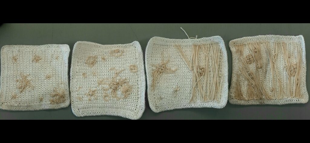
This is a culminative picture of my project over the last 2 semesters, I have some more detailed and labelled pictures below.
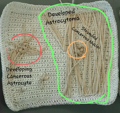
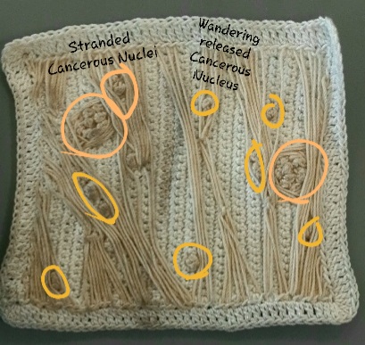
This is the final image of developed Astrocytoma. In this image the branches have become long, thin, and tangled with one another. There is also evidence of the burst cell membrane because of the presence of all the free floating Cancerous Nucleus. There are a few cells that are unburst as well which I have also marked.
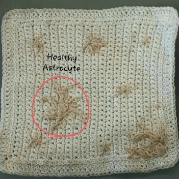
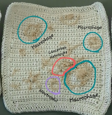
Calyssa did her project on the development of Astrocytoma. Astrocytoma is a type of cancer that can occur in the brain. She described that Astrocytes are blood-brain barriers that are important for determining what is coming in and out of the brain. She crocheted and focused on Glioblastoma. Symptoms of astrocytoma are nausea, muscle weakness, memory issues, seizures, headaches, and difficulty sleeping. Astrocytoma usually originates in the brain and not other parts of the body. Calyssa crocheted square pieces of “brain matter” in different stages showing the development of an astrocytoma. The first stage shows a healthy astrocyte in the grey brain matter, it also has a concise and a nucleus. In the second square, she displayed a cell that has become cancerous. There is also a macrophage and neutrophils showing that they came to take care of the cancerous astrocyte. The third square, it is showing that the macrophages failed in their attempt to destroy the cancerous cell. At this point the cancerous astrocyte starts to develop the long spindly limbs, then the limbs begin to knot with other cancerous cells. In the last stage, the limbs and branches have become very long and tangled and knots have occurred. Overall Calyssa did a great job it was a very unique way to see how brain cancer occurs when typically it is something we cannot see.