
My project is about hemophilia, which is a disease where an individual doesn’t have all the right clotting factors so they cannot form blood clots quickly or at all. I created two models, one which shows an individual’s vessel that formed a blood clot. It shows platelets, clotting factors, and fibrin which holds it all together. The other vessel is a vessel of someone that has hemophilia where all the clotting factors are not present, which means that the platelets cannot stick together right and the fibrin does not bind everything together.
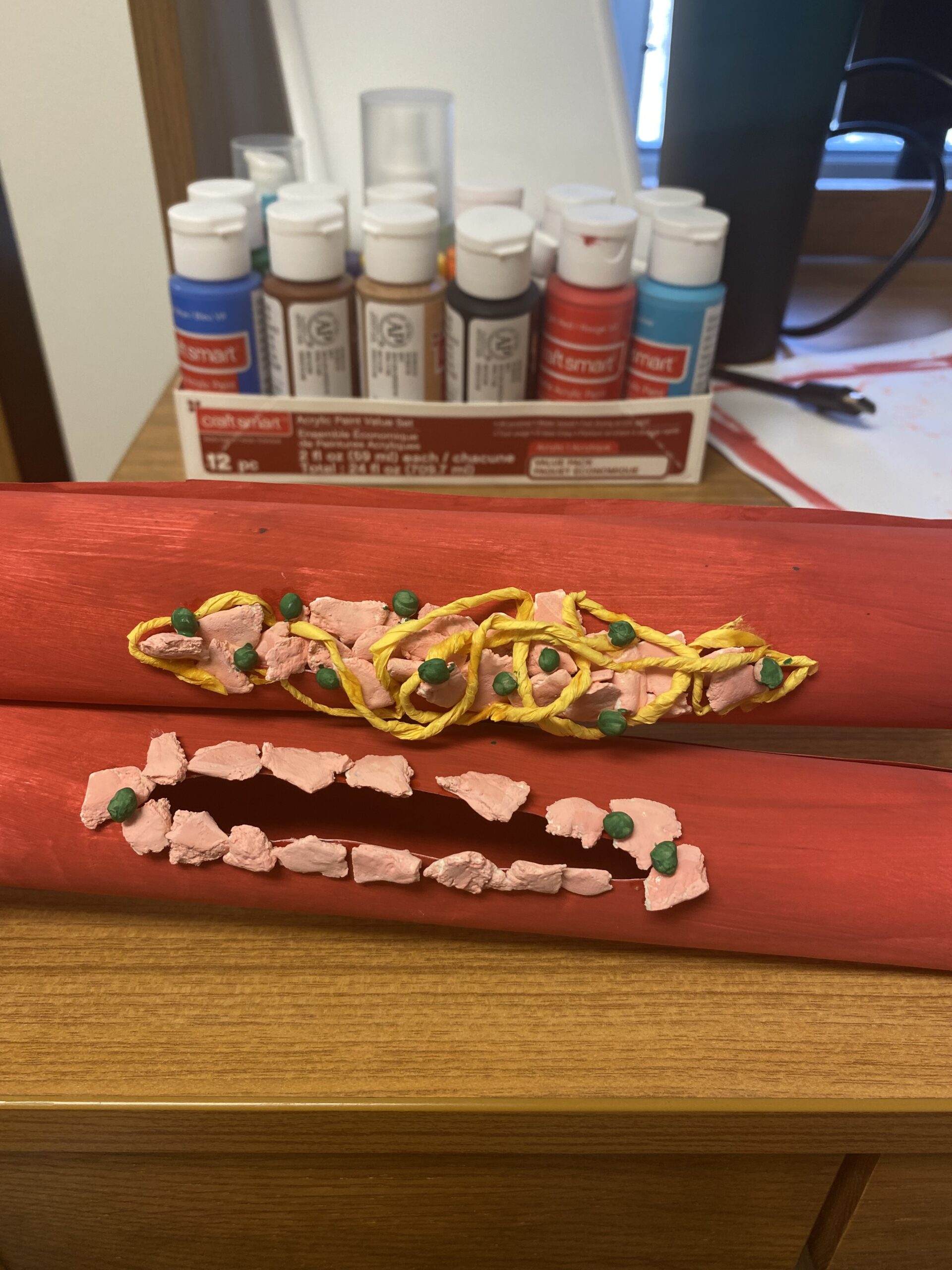
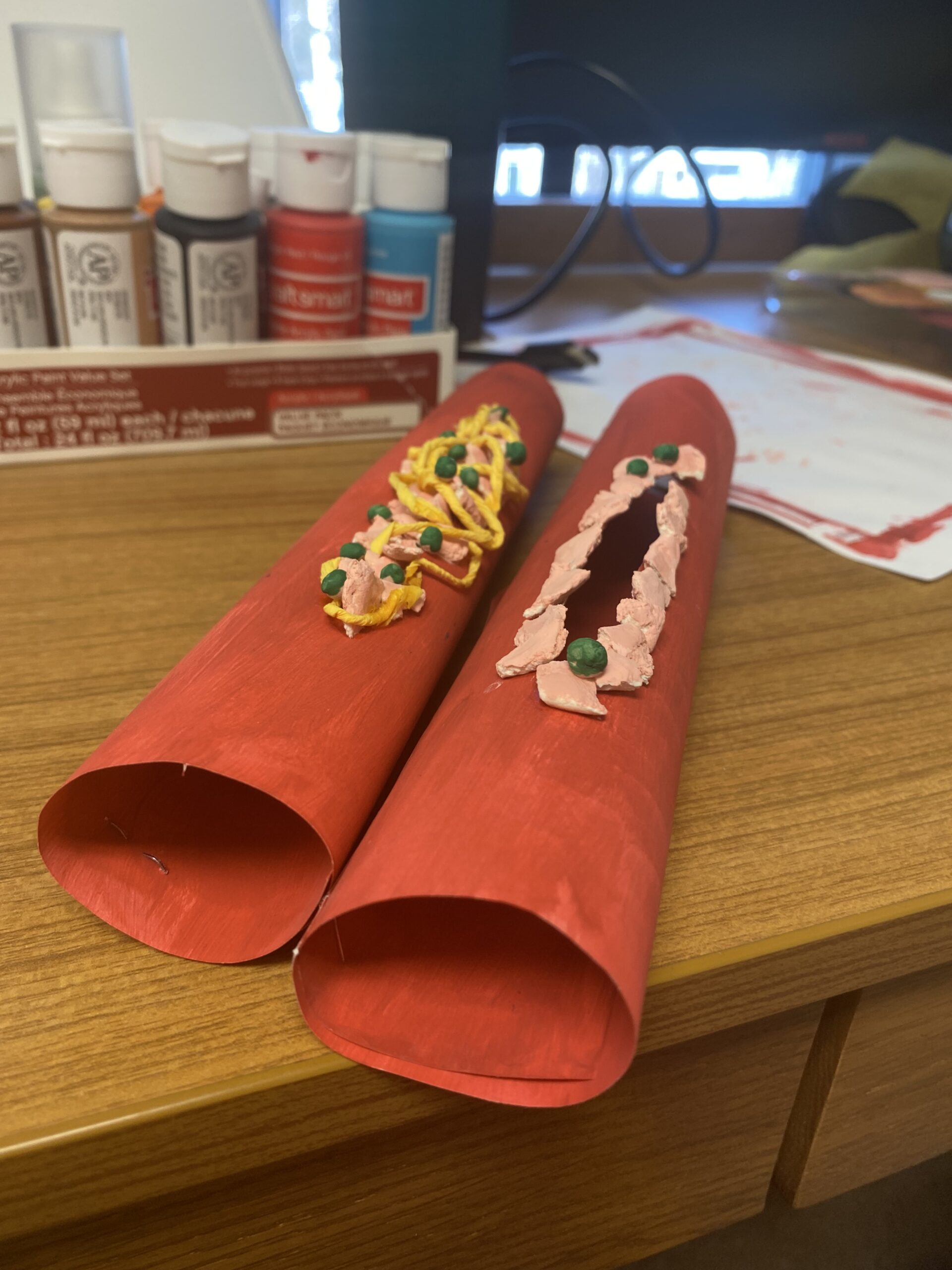
This model artfully depicts the differences in blood clotting in a normal body versus in someone with hemophilia. Both models depict the first step of blood clotting which is vasoconstriction. Vasoconstriction is muscles contraction to limit the blood flow to the affected area. This action is the same in people with and without hemophilia. The second step, platelets sticking to the vessel, are depicted on both models with small green pellets. Someone without hemophilia will then go through a clotting factor cascade. All the clotting factors work together to trigger clotting fibers and to help platelets stick together. In the first model, you can see the platelets and fibrin helping to hold a clot together.
Someone with hemophilia either has too low of a level or is missing a clotting factor. This halts the process of stopping or making the person extremely slow to clot. In the second model, you can see the process has been halted, leaving an open wound. This condition can be very dangerous because it can make small injuries fatal. People may bleed excessively internally or bleed in their brain both of which can be fatal. However, it is easily treatable by administering concentrated forms of clotting factors VII and IX to make up for what the person with hemophilia is missing.