
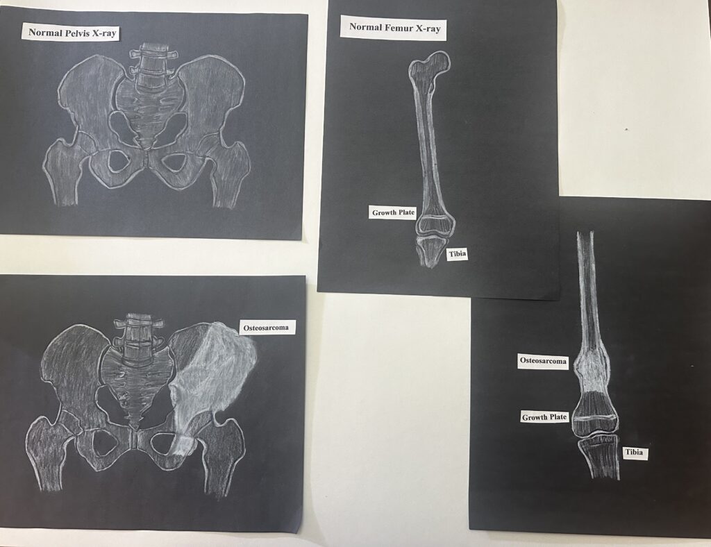
For my STEAM project, I am focusing on cells that comprise bone tissue and relating these cells to the development of osteosarcoma. In my art project you will find femur and pelvis images that are healthy and an image of each with osteosarcoma. Totaling in four images to look like an x-ray, as this is the most common tool used to diagnose osteosarcoma.
Amy’s STEAM project about bone tissue development, remodeling, and osteosarcomas provides easily understandable information about how bone tissue and remodeling present in osteogenic sarcoma, or bone cancer. She explains how bone develops from osteoblasts and bone matrix that calcifies into osteocytes which are the main component of bone tissue. Amy also explains the process of bone remodeling through osteoblast and osteoclast activity.
The visual presentation of osteosarcoma x-rays depicts the most common sites that osteosarcomas develop. Primary osteosarcomas are more common in children and teenagers and tend to develop in the long bones of the appendicular skeleton, most commonly on the distal aspect of the bone in the growth plates. Secondary osteosarcomas are more common within older adults (50+) and present in the pelvis, generally with late symptom onset.
The cause of osteosarcomas is not definitively known but are thought to involve mesenchymal cells or osteoblast precursor cells. Mutations in cells that form bones creates mutations in tissues that form into tumors and cause bones to be porous and brittle. It is common to have bone fractures and breaks of unknown origin with osteosarcomas. Symptoms of possible osteosarcoma include swelling and lumps on bones, joint and bone pain, and soreness. are often diagnosed through x-ray imaging and are treated with tumor removal as they have an aggressive clinical course. Appendicular osteosarcomas generally have a good prognosis and less is known about pelvic osteosarcoma prognosis.