

Abstract
For my STEAM project, I focused on the objective statement “Identify the various components and key structures of the nervous system.” To do so I decided to build a model of the human brain showing the cerebrum, cerebellum, and brain stem. I then focused on the areas involved in language comprehension for dyslexic and non-dyslexic people. This involved further research on the subject. Then I modeled the active areas of reading comprehension of either group (Dyslexic and non-dyslexic) on my model.
Process
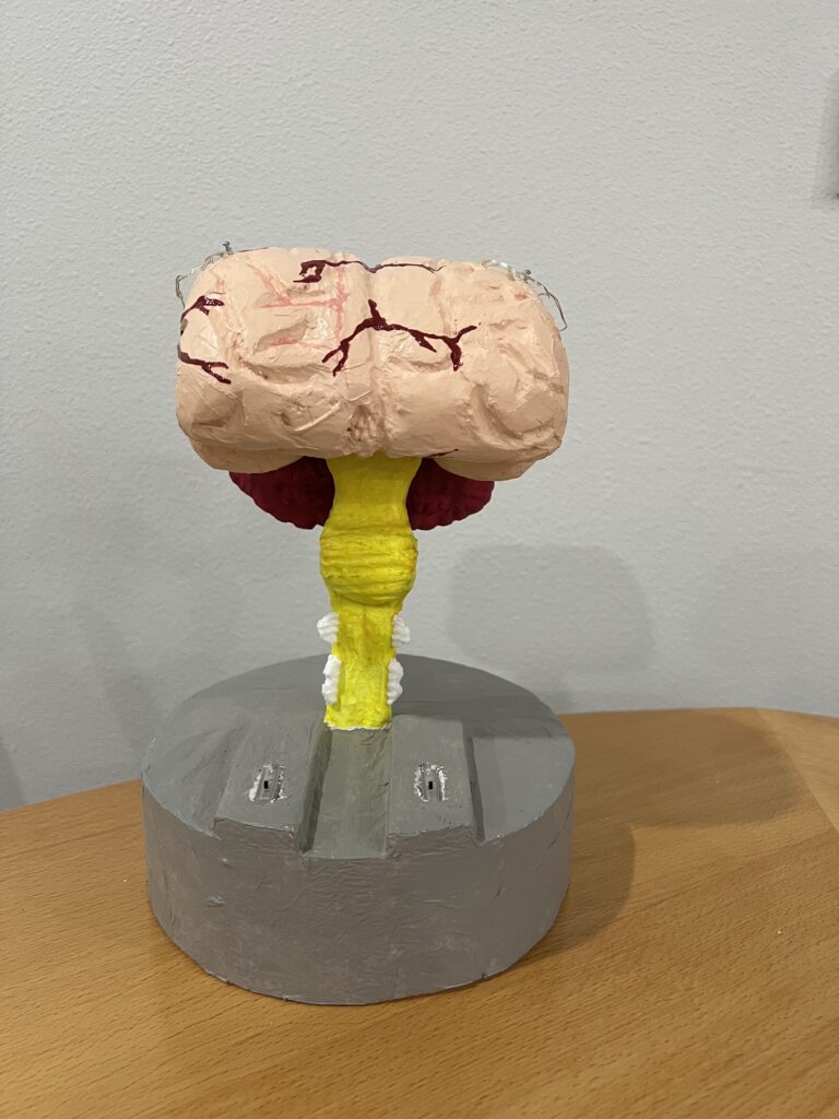
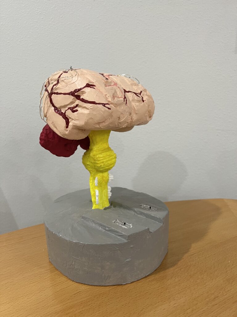
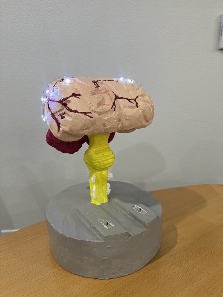
To construct the brain model, I used the resources provided by BioMedical Art on YouTube for directions. This consists of three videos; their citations are on the resource page. The main model consists of Styrofoam carved to the shape of the cerebrum (BioMedical Art, Sep. 5, 2018), cerebellum (BioMedical Art, Sep. 6, 2018a), and brain stem (BioMedical Art, Sep. 6, 2018b). I then covered styrofoam in a collage of tissue paper and painted over the tissue once dry. To model the activity of the brain during language comprehension I used LED string lights (sometimes referred to as fairy lights). These lights were wired through the styrofoam model and connected to two switches. The left switch turns on the area of the brain connected to dyslexia and the right switch represents the brain activity of a non-dyslexic person. To find the exact areas to model I used MRI (magnetic resonance imaging) photos from NeuroImage’s article on language comprehension in dyslexic children (Van der Mark, et al., 2011).
Research
Dyslexia is a “severe reading disorder, which is characterized by dysfluent reading and impaired automaticity of visual word processing” (Van der Mark, et al., 2011). The condition is due to neurological abnormalities in language comprehension areas such as the “left occipito-temporal cortex (OTC), temporo-parietal cortex (TPC), and frontal area” (Hernández-Vásquez, R., el al., 2023). Studies involving electrophysiological evidence (used in reflecting the electrical activity of cortical neurons) show that the problem lies in functional and structural problems of the left hemisphere “including reduced grey matter volume in the left TPC, decreased white matter connectivity between reading networks, and hypoactivation of the left OTC and TPC” (Hernández-Vásquez, R., el al., 2023). The study cited above by the Iranian Journal of Psychiatry supports the conclusion of decreased connectivity in dyslexic brains in the left occipitotemporal system (Van der Mark, et al., 2011).
In my model, I showed areas of the occipitotemporal cortex, and the temporoparietal cortex active in normal brains and dyslexic brains during language processing. I used MRI images in NeuroImage’s study in the process of mapping my brain model (Van der Mark, et al., 2011). My mapping shows normal brain activity in both the right and left hemispheres, which is indicated by the occipitotemporal cortex, and the temporoparietal cortex of either hemisphere being active (lit up with the LED string light) with emphasis on the left. In my model, dyslexic brains show no activity in the right hemisphere, and reduced activity in the left hemisphere, especially in the temporoparietal cortex. The indications of my mapping are supported by the findings of electrophysiological studies in that they indicate “reduced connectivity between occipital to inferior-temporal areas, as well as between left to right temporal regions.” (Hernández-Vásquez, R., el al., 2023). This conclusion is indicated by my model.
References
BioMedical Art. (2018, September 5). How to make Brain Cerebrum Model | 3d thermocol/Styrofoam Carving [Video]. YouTube. YouTube. Retrieved November 21, 2023, from https://www.youtube.com/watch?v=wgxUFs3aDR8.
BioMedical Art. (2018a, September 6). How to make Brain Cerebellum Model |3d thermocol/Styrofoam carving [Video]. YouTube. YouTube. Retrieved November 21, 2023, from https://www.youtube.com/watch?v=dD1cxFLScMo.
BioMedical Art. (2018b, Spetember 6). How to make Brain Stem Model | 3d thermocol/Styrofoam carving [Video]. YouTube. Retrieved November 21, 2023, from https://www.youtube.com/watch?v=dbb3LmkJqVQ.
Hernández-Vásquez, R., Córdova García, U., Boy Barreto, A. M., Rodriguez Rojas, M. L., Ponce-Meza, J., & Saavedra-López, M. (2023). An Overview on Electrophysiological and Neuroimaging Findings in Dyslexia. Iranian Journal of Psychiatry, 18(4), 503–509.
Van der Mark, S., Klaver, P., Bucher, K., Maurer, U., Schulz, E., Brem, S., Martin, E., & Brandeis, D. (2011). The left occipitotemporal system in reading: Disruption of focal fMRI connectivity to left inferior frontal and inferior parietal language areas in children with dyslexia. NeuroImage, 54(3), 2426–2436. https://doi.org/10.1016/j.neuroimage.2010.10.002
The project written by Asher McGlinchy is on brain mapping for language comprehension of dyslexic and non-dyslexic brains. The objective he covered is “identify the various components and key structures of the nervous system”. For his art project, he constructed a brain model showing the areas of the brain that are used for language comprehension in dyslexic and non-dyslexic people.
Dyslexia is a reading disorder that can impede reading and visual word processing. This condition is due to abnormalities in the language comprehension areas of the brain. The main functional and structural problems that cause this disorder are found in the left hemisphere, including reduced gray matter volume and decreased white matter. In a non-dyslexic brain, both hemispheres of the brain function properly and can be used for language comprehension. In dyslexic brains, there is no activity in the right hemisphere and reduced activity in the left hemisphere. He used his model to convey this by using lights that lit up to show what part of the brain is being used in dyslexic and non-dyslexic brains.