
Bio F111
Steam Project
20/11/2023
Have you wondered how scientists have bioengineered healthy skeletal muscle tissues to repair or replace acute injuries?
Scientists are finding ways for diseased or damaged muscle tissue to build upon healthy muscle tissues to help with muscle repairs and replacements; from C. Handschin, scientists are using a piece of tissues to engineer cell-based muscle tissue to revert the functional loss of muscle tissue caused by a disease or trauma. Even though muscle tissue can be bioengineered using precursor cells, some limitations remain. The fundamental limitations are the cellular pathways crucial to muscle tissue regeneration. The regeneration process needs external stimuli to orchestrate the regeneration process after physiological wear and muscle trauma. Still, they are the most crucial part of the aging process and maintain the niche of the stem cells, which are responsible for long-term muscle function.
The objective is to see the interaction between the muscles and the skeletal muscle system.
As many athletes continue to play their sports accordingly, they receive injuries and tears from an impacted fall or torn-up skeletal muscle tissue. They now have issues with playing their sport and their everyday life. Now, they must talk to the family care physician to help them understand the injury and the possible results that can help them heal. However, what could be more engaging is how scientists use models to investigate and develop therapeutic strategies for muscle injuries and provide studies for severe neuromuscular and musculoskeletal disorders.
To understand how it works, we need to know the components and functions of the skeletal muscle and why it is essential to know why it is the most abundant human tissue. The functions include movement, generation, posture maintenance, storage of the nutrient’s metabolites, soft tissue support, and maintaining the body’s temperature homeostasis. The cellular units of skeletal muscle are the muscle fibers(myofibers), long cylindrical multinucleated cells containing the contractile apparatus, which are densely packed along a force axis. From our Biology F111 class lesson, we learned that skeletal muscle contractions and force productions are made when actin and myosin filaments are arranged in units called sarcomeres. Then, the skeletal muscle is innervated by the motor neurons that interact with the neuromuscular junctions. The neurotransmitter acetylcholine is released in synapses, activating the signaling pathways, ultimately leading to muscle contraction.
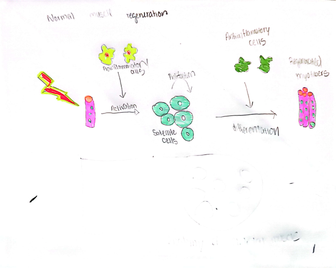
To help it contract eventually, some scientists are finding ways to have the regenerative tissue help the tissue heal up. To base the engineering process, we need to know the regenerative tissue phase steps to know how the tissue can be repaired. The phases are to rule out the tissue injury to see what it is, how the inflammatory response is, the activation, and then the matured and remodeled tissue. The model I painted shows the process of skeletal muscle regeneration and the process many scientists use to engineer the skeletal muscle to act as an implant in place of the torn or damaged muscle. The first depiction of my image is when the muscle is damaged. The mononucleated quiescent cells are stimulated to divide to fuse with existing muscle fibers to regenerate and repair damaged fibers. The skeletal muscle fibers themselves cannot divide; however, the muscle fibers can lay down new proteins and enlarge.
Handschin, C., Mortezavi, A., Plock, J., & Eberli, D. (2015, October 22). External physical and biochemical stimulation to enhance skeletal muscle bioengineering. Sciencedirect. https://doi-org.uaf.idm.oclc.org/10.1016/j.addr.2014.10.021
Jiang, Y., Torun, T., Maffioletti, S. M., Serio, A., & Tedesco, F. S. (2022). Bioengineering human skeletal muscle models: Recent advances, current challenges and future perspectives. Experimental Cell Research. https://doi-org.uaf.idm.oclc.org/10.1016/j.yexcr.2022.113133
Paxton, S., Peckham, M., & Knibbs, A. (1970, January 1). The Leeds Histology Guide. Home: The Histology Guide. https://www.histology.leeds.ac.uk/tissue_types/muscle/muscle_regeneration.php#:~:text=When%20the%20muscle%20is%20damaged,protein%20and%20enlarge%20(hypertrophy).
University, W. F. (2023). Engineering muscle implants. Wake Forest University School of Medicine. https://school.wakehealth.edu/research/institutes-and-centers/wake-forest-institute-for-regenerative-medicine/research/military-applications/engineering-muscle-implants
Wang, W., Xin-Hua, Stephenson, L. L., Khiabani, K. T., & Zamboni, W. A. (2009, February). Ischemia-reperfusion–induced apoptotic endothelial cells isolated from … Ischemia-Reperfusion–Induced Apoptotic Endothelial Cells Isolated from Rat Skeletal Muscle. https://www.semanticscholar.org/paper/Ischemia-Reperfusion%E2%80%93Induced-Apoptotic-Endothelial-Wang-Fang/abb70b22e90b87fda57fd97c49d9b69b92e58551
Ziemkiewicz, N., Hilliard, G., Pullen, N. A., & Garg, K. (2021, March 23). The role of innate and adaptive immune cells in skeletal muscle regeneration. MDPI. https://www.mdpi.com/1422-0067/22/6/3265
ps. why is the apa citations not indenting the second and the rest of the lines?
This project explores cutting-edge bioengineering strategies to repair and replace damaged skeletal muscle tissues resulting from acute injuries. The study focuses on the intricate interplay between muscles and the skeletal muscle system, which is crucial for athletes facing injuries impacting their sports and daily lives. By dissecting the components and functions of skeletal muscle, the project elucidates why it is the most abundant human tissue, emphasizing movement, posture maintenance, and temperature homeostasis. The regenerative tissue phase, encompassing injury recognition, inflammatory response, activation, and tissue maturation, is a foundation for engineering skeletal muscle implants. The depicted model illustrates the sequential steps in skeletal muscle regeneration, highlighting the potential of engineered tissues to replace damaged muscle effectively.
This project investigates how healthy skeletal muscle tissue can repair or replace acute injuries. This project goes beyond the traditional methods of healing and seeks to reason using an alternative method of injury or disease treatment. The project’s focus on sport-related injuries and muscular disease outlines a necessity for a faster healing process. As described in the written statement, utilizing a sample of tissue to rewire healthy skeletal muscle to repair the functional loss caused by injury or disease proved more beneficial on time constraints than traditional methods for healing. Although, there are a few downsides to this method with the involvement of cellular pathways. With the damage and aging of muscles, the cellular pathways begin to require external stimuli to initiate the regenerative process. As these cellular pathways are vital to all muscular repair, they are required for long-term muscular function.
This treatment option is viable due to the intermuscular interactions between the muscle components during contraction. Contraction occurs when the actin and myosin proteins are aligned within sarcomeres. Motor neurons that interact with neuromuscular junctions innervate the muscle by releasing acetylcholine from their synapses. This acetylcholine activates the signaling pathways and thus begins contraction. Scientists use this process of contraction and compare it to the regenerative phases of tissue repair to pinpoint where the sample skeletal muscle tissue would be best placed. By analyzing the contraction process, the project outlines how using skeletal muscle to regenerate damage is a viable treatment option for skeletal muscle wear and tear.