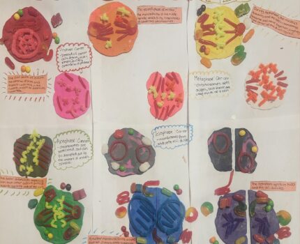
I chose to represent the course objective of describing the stages of mitosis through a clay and candy diagram. “Perhaps the most amazing thing about mitosis is its precision, a feature that has intrigued biologists since Walter Flemming first described chromosomes in the late 1800’s” (Paweletz, 2001). Mitosis is a process within the cell cycle where a cell replicates their chromosomes, equalizes the outcome, then produces two identical nuclei in preparation for cell division. In the end, Mitosis results with two identical daughter cells with uniform genomes. For example, mitosis could be thought of as a school filled with students of all different grades yet have to follow the same guidelines. This school would not want one class to receive loads of information, while the other classes received little to no information on the guidelines, almost like allowing the senior class free roam of a school. If a mistake like that arose, the cells may have difficulties following protocol. Thankfully the cells within our bodies have great systems to copy cells without major errors. Mistakes do happen, and the body ultimately suffers (Gilchrist, 2023).
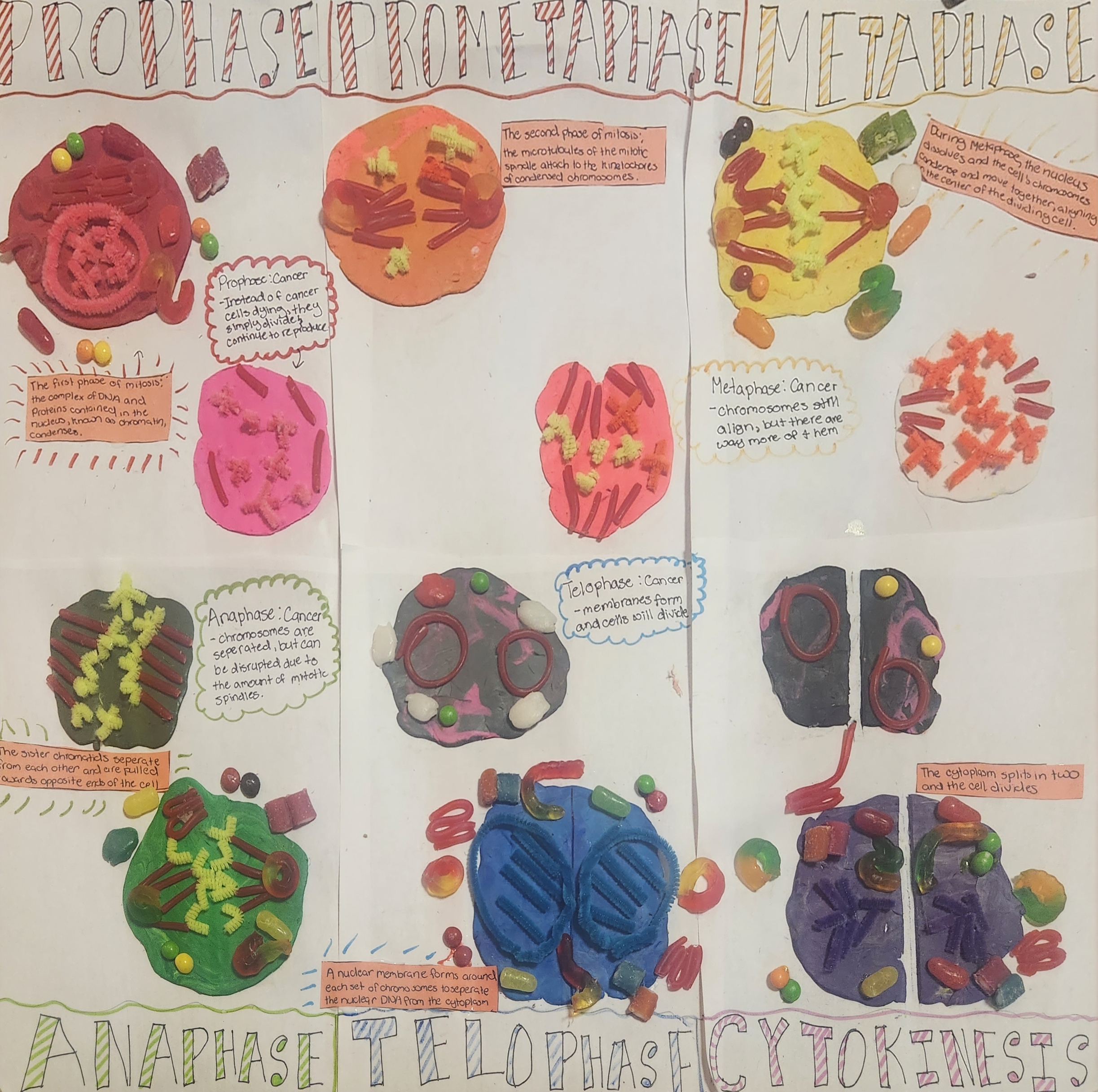
The mitosis cycle results in five crucial stages, each with a primary role to help our bodies contain equilibrium. These stages include Prophase, Prometaphase, Metaphase, Anaphase, Telophase, and Cytokinesis. To begin the cycle, the cells undergo prophase, where the complex of DNA and proteins within the nucleus (known as chromatin) condenses. The spindle begins its formation as the two centriole pairs move to opposite poles, and the microtubules start to polymerize from the duplicated centrosomes. Next, the cells begin prometaphase, which begins with the abrupt fragmentation of the nuclear envelope into small vesicles that may then divide between the two daughter cells. Also, the microtubules of the mitotic spindle attach to the kinetochores of the condensed chromosomes. Following this stage comes metaphase where the chromosomes are lining up in the center of the cell in preparation for the next stage, anaphase. Anaphase is where two crucial movements occur within the cell. First, the kinetochore microtubules shorten then move towards their spindle poles. Then the spindles separate as the non-kinetochore would move past one another in a latter movement. These sister chromatids are pulled to opposite sides of the cell towards their designated spindle. Lastly the cycle concludes the system with telophase and cytokinesis. Telophase is where the chromosomes have reached the poles, the nuclear envelope has reformed, and the chromosomes begin to decondense once again. Also, as the nuclear envelopes reform, they end up creating a wall around each set of chromosomes to help separate the nuclear DNA from the cytoplasm in preparation for cytokinesis. Cytokinesis is well known as the stage that divides the cytoplasm into two identical daughter cells. This is the process of mitosis in a regular, healthy cell (Connor, 2014).
On the contrary, if a cell undergoes mitosis with a cancerous cell, the number of chromosomes is exponentially greater than those in a regular, healthy cell. The cycle still would begin with prophase, and instead of the cancer cells dying they instead divide and reproduce. Within Metaphase, the chromosomes are still going to align at the center of the cell; However, there are way more of the chromosomes present. Next in anaphase, the chromosomes are separated, but can still be disrupted due to the amount of mitotic spindles present. Finally, telophase, the membranes will continue to form, and the cells will continue to divide forever. Now remember when I stated earlier how one mistake could have long lasting effects on a person’s cells, well if a mistake occurs within the genetic makeup of the cells, cancerous cells arise. The Mitosis cycle occurs infinitely, meaning that even with infected cells the cycle just keeps going on forever. Cancerous cells never die, as they utilize telomerase to add more telomerase sections in a cell’s DNA strands prior to replication (Mercadante, Anup, 2023). Cancer cells are ultimately cells that have gone wrong, which originate within the body’s tissues and as they grow and divide, so does their resistance to the cell’s natural defenders. Cancerous cells can evade apoptosis and continue to metastasize. The mutations within the genes can cause cancer by accelerating cell division or inhibiting apoptosis and cellular arrest. The more that cancerous cells divide, the stronger and more unstoppable they become, making cancerous cells an unstoppable villain within the cycle of life (Nature Education, 2014).
I represented the objective of mitosis with a poster diagram of the cell cycle. I chose to utilize candy and clay because of a memory from my last year of middle school where we had to create the cell cycle with candy. I thought that it would be fun to go down memory lane and dive deeper into the cycles of mitosis. I ended up utilizing the clay as a base for each phase of mitosis, and the candy as both the spindles and the exterior attributes of a typical cell. What I mean by this is within the regular, healthy cells, I incorporated attributes like mitochondria, vacuoles, centrioles, ribosomes, Golgi apparatus, and endoplasmic reticulum, along with the actual phase of course. All these attributes of the cell cycle were represented by a different piece of candy. The mitochondria are represented by mike & nikes, the vacuoles by airheads, the centrioles by sweet tart ropes, the Golgi apparatus by a gummy worm, and the endoplasmic reticulum by a twizzler. All together forming this colorful diagram of the mitosis cell cycle. I honestly thought that this would take me far less time to do, but in the end took me about two to three days to complete. I was ultimately surprised to see that both cancerous and healthy cells undergo the same phases of mitosis, with the only difference being that cancerous cells never die and continue to reproduce and form exponentially larger amounts of chromosomes, disrupting the equilibrium of a person’s body.
Citations:
Connor, C. O. (2014). Mitosis and Cell Division. Nature news. https://www.nature.com/scitable/topicpage/mitosis-and-cell-division-205/
Gilchrist, D. A. (n.d.). Mitosis. Genome.gov. https://www.genome.gov/genetics-glossary/Mitosis
Mercadante, A. A., & Kasi, A. (2023, August 14). Genetics, cancer cell cycle phases – statpearls – NCBI bookshelf. National Library of Medicine. https://www.ncbi.nlm.nih.gov/books/NBK563158/
Nature Publishing Group. (2014). Cell Division and Cancer. Nature news. https://www.nature.com/scitable/topicpage/cell-division-and-cancer-14046590/
Gabreall’s project explains the process of Mitosis. He then explains how Mitosis is different in cancerous cells. In a cancerous cell the chromosomes still line up during the Metaphase process, however, there are more chromosomes than in a healthy cell. In Anaphase, the chromosomes separate like they do in a healthy cell, but this time because there are more spindles present from more chromosomes, disruption can still take place. In Telophase, the new membranes form as they should, but the cells will continue to divide forever. This continual division just keeps the cancer replicating and spreading.
Gabreall’s explanation helped me get a clear picture of why cancer can be so deadly. Because it just keeps copying the cancerous genetic information, it is making new cancer and introducing it to the body. The body is doing what it should in Mitosis, but it is instead making deadly cells to harm itself. Since he explained that the more the cells divide, the stronger the cancer becomes, I got a clear idea of why treatments sometimes don’t work, and why sometimes doctor’s report cancer has spread too far to treat. It is ironic that the body doing what it should actually makes the thing harming it stronger.
The poster was creative and colorful. The poster gave me a visual that helped fill in some more information that just the paper did. I think a key would have been helpful to explain what each piece of candy corresponded to as parts of the cell.