


The Structure of the Knee Joint and Ligament Injuries
The knee joint is a complex and unique structure, classified as a synovial joint but organized differently from any other joint in the body. My project will be covering the course objective “know the structure of the knee joint and compare this joint type to other 5 joint types” and will focus on the anterior cruciate ligament (ACL) and posterior cruciate ligament (PCL).
The knee is classified as a synovial joint, meaning that the bones forming the joint are not directly connected to each other via connective tissue or cartilage. Instead, there is a fluid-filled cavity between the two bones that allows them to contact each other and articulate without being directly connected. Synovial joints are the most common type of joint found in the body, and there are six different types: pivot, hinge, condyloid, saddle, plane, and ball-and socket-joints. The knee is classified as a “hinge joint,” which means that the convex end of one bone articulates with the concave end of the adjoining bone. The structure of a hinge synovial joint is very different from the types of fibrous (sutures, syndesmoses, and gomphoses) and cartilaginous (synchondroses and symphyses) joints. Due to their structure, fibrous and cartilaginous joints do not allow for nearly as much movement as the synovial joint of the knee does.
Although most joints in the body are synovial joints, the knee joint is the largest and most unique in that it is comprised of three different joints and includes an additional bone called the patella (or kneecap) which is incorporated into the quadriceps femoris muscle. Those three different joints are the femoropatellar joint (between the patella and the distal femur), the medial tibiofemoral joint (between the medial condyles of the femur and tibia) and lateral tibiofemoral joint (between the lateral condyles of the femur and tibia). Between the articulating surfaces of the femur and tibia are the medial meniscus and lateral meniscus, two thin fibrocartilage structures that attach to the condyles of the tibia. These menisci provide padding between the tibia and the femur, in the gap between the rounded femoral condyles and the flattened tibial condyles.
There are also several ligaments that support the knee joint and allow it to function, including the fibular collateral ligament which connects the fibula to the femur, and the tibial collateral ligament which connects the tibia to the femur. These two collateral ligaments are taut when the knee is fully extended, which stabilizes and supports the knee and helps to prevent side-to-side or rotational movements between the femur and tibia. They are both located outside of the articular capsule at the sides of the knee.
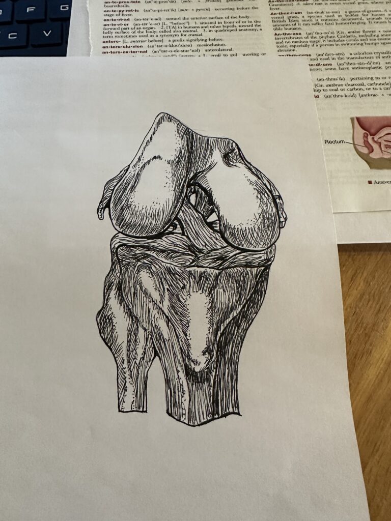
There are two ligaments inside the articular capsule of the knee that help to prevent hyperextension of the knee: the anterior cruciate ligament (ACL) and the posterior cruciate ligament (PCL). The ACL is attached to the anterior side of the tibial intercondylar eminence, which is the roughened area between the tibial condyles. Conversely, the PCL is attached to the posterior side of the tibial intercondylar eminence. Each of these ligaments runs diagonally upward to attach to the femur, passing over each other in an X-like position as they do so. The PCL is stronger than the ACL, and its position holds the joint together and prevents the femur from sliding anteriorly (forward) off the top of the tibia. It supports the knee during flexion and weight-bearing. The ACL restrains forward migration of its tibial attachment and prevents hyperextension by becoming taut when the knee is extended to inhibit further extension (Fuss, 1991).
Knee injuries are common in athletes and can result in partial or complete tears of the ACL or PCL. However, PCL tears are much less common than ACL tears due to the size and strength of the PCL comparatively (Wang, 2018). Diagnosing a cruciate ligament injury can be done through Magnetic Resonance Imaging, which can determine the extent of the injury and what treatment is required to repair it (Sultana, 2023). Treating an ACL or PCL tear may involve reconstruction surgery to reattach or reconnect the ligament. This often results in a full recovery with complete regaining of abilities, but this is not always the case.
Specifically with the PCL, surgical reconstruction may only be recommended in acute injuries that result in severe posterior tibial movement and instability of the knee, or if there is damage to other structures in the knee in addition to the PCL (Cox, 2022). People with catastrophic PCL damage can still have functional mobility of the knee with non-operative treatment, meaning that they do not undergo surgery to reconstruct the PCL. The body can adapt to the PCL injury and continue to function almost normally, though they will have some instability in the knee and may be at risk for further damage to structures in the knee (such as meniscus tears) as time progresses (Wang, 2018).
The knee joint is unique compared to other joints in the body and stands out as the largest and most complex synovial joint. There are four main ligaments that support knee mobility, stability, and function: the fibular and tibial collateral ligaments (outside of the articular capsule) and the anterior and posterior cruciate ligaments (inside of the articular capsule). While ACL and PCL injuries are common, treatment can vary depending on the severity of the damage and the affect the injury has on mobility and function of the knee.
References
Cox CF, Graefe SB, Bordoni B. Anatomy, Bony Pelvis and Lower Limb: Knee Posterior Cruciate Ligament. [Updated 2022 Jul 25]. In: StatPearls [Internet]. Treasure Island (FL): StatPearls Publishing; 2023 Jan-. Available from: https://www.ncbi.nlm.nih.gov/books/NBK535416/
Fuss FK. The restraining function of the cruciate ligaments on hyperextension and hyperflexion of the human knee joint. Anat Rec. 1991 Jun;230(2):283-9. doi: 10.1002/ar.1092300217. PMID: 1867405.
Sultana N, Shirin M, Jabeen S, Faruque MA, Sarkar SK, Nag UK, Nabi S. Diagnostic Accuracy of Magnetic Resonance Imaging in Evaluation of Anterior Cruciate Ligament Tear. Mymensingh Med J. 2023 Jan;32(1):200-206. PMID: 36594321.
Wang, S. H., Chien, W. C., Chung, C. H., Wang, Y. C., Lin, L. C., & Pan, R. Y. (2018). Long-term results of posterior cruciate ligament tear with or without reconstruction: A nationwide, population-based cohort study. PloS one, 13(10), e0205118. https://doi.org/10.1371/journal.pone.0205118
In CJ Pensabene’s project, she created a visual art project on the Structure of the Knee Joint. More specifically, she has chosen to dive into common ligament injuries like with the anterior cruciate ligament (ACL) and the posterior cruciate ligament (PCL). The course objective covered by this project is to “know the structure of the knee joint and compare this joint type to other 5 joint types”.
CJ focuses primarily on the Posterior Cruciate Ligament, and the Anterior Cruciate Ligament, while also presenting a complete examination of the knee joint’s structure. The project goes deep into the knee joint’s complicated structure, for instance that “the knee is classified as a a synovial joint which means that they are not directly connected to each other by connective tissues or cartilage. Instead, there is a fluid-filled cavity between the two bones that allows them to contact each other and articulate without being directly connected.” CJ also talks about the entirety of the knee joint going over how it functions, and importantly, how the ACL and PCL prevent hyperextending the knee. She also talks about other common injuries for athletes and dives even deeper into the ACL and PCL, like tears, how to diagnosis through medical imaging, and how to treat the tears through reconstruction surgery.
For Ms. Pensabene’s study, she covered a very complex subject that can be easily understood by others. Her research will give anyone who reads it a much better understanding of the knee joint and how critical the ACL and PCL are, along with injuries to these ligaments. The essay provided is clear and concise and it gives readers an opportunity to better comprehend the many parts of the knee joint’s unique structure.