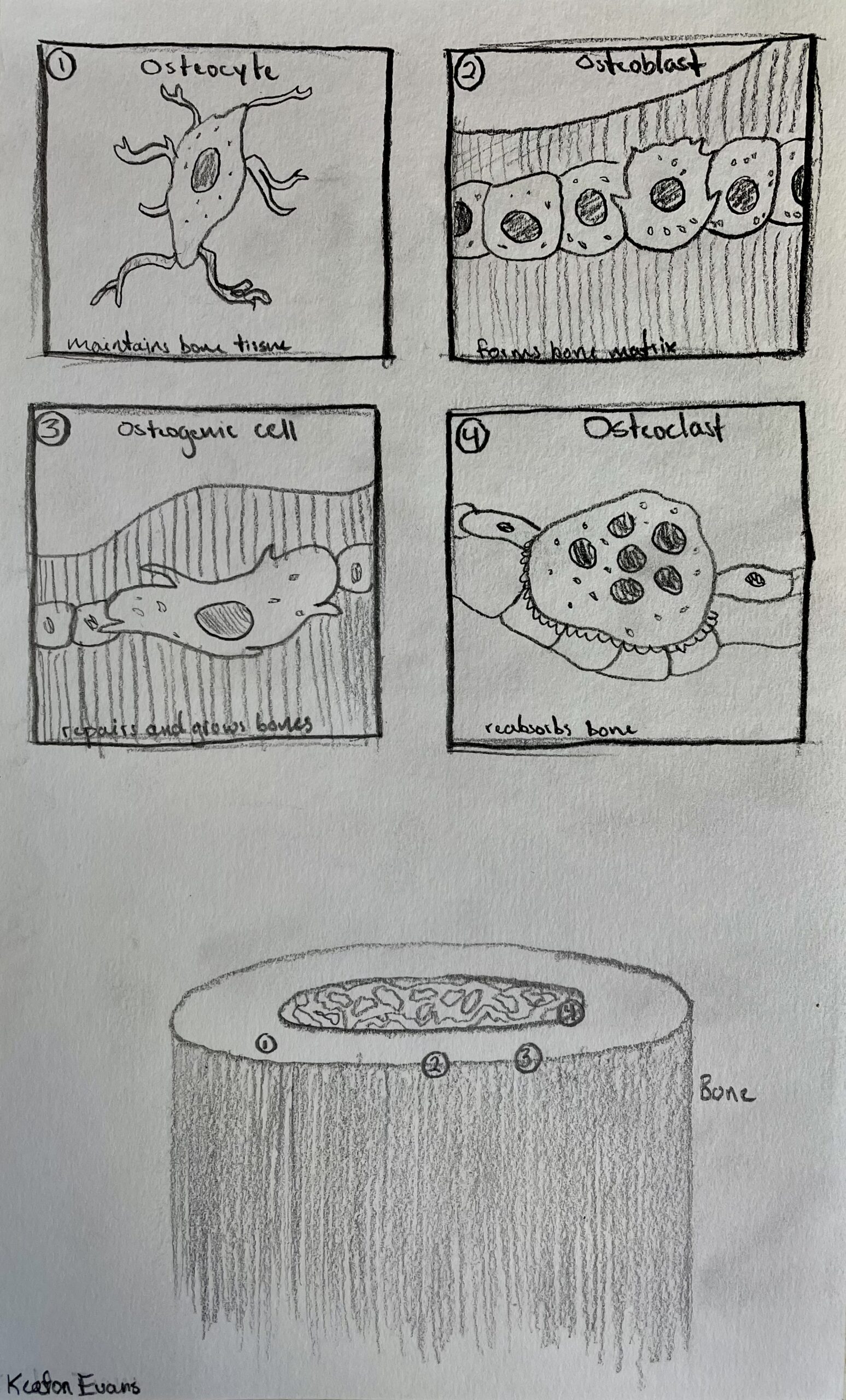
The course objective I am going to cover in this STEAM is “Identify the 4 cells that comprise bone tissue.” Artistically, I am going to use graphite drawings to show each cell and their respective details. The skeletal system interested me the most during this class and I am excited to share what I found in my research. The four cells that comprise bone tissue are: 1.) Osteoblasts, 2.) Osteocytes, 3.) Osteoclasts, and 4.) Osteogenic cells. Through my research I was able to discover more about each one.
Osteoblasts serve a critical role within bone tissue as they regulate osteoclasts and depose of bone matrix. Osteoblasts themselves are, as one scholarly article put it, “mononuclear, not terminally differentiated, specialized cells” (Caetano-Lopes, 1970, p. 104) and that they “form tight junctions with adjacent osteoblasts and have regions of the plasma membrane specialized in vesicular trafficking and secretion” (Caetano-Lopes, 1970, p. 104).
Osteocytes are by far the most abundant cell type found in bone. As one article puts it, (referring to the function of an Osteocyte), [an Osteocyte] “is the principal cell type responsible for integrating the mechanical and chemical signals that govern modeling and remodeling and control the onset of both bone resorption and formation” (Schaffler, 2013, p. 6). Basically, the cell needed to signal Osteoblasts and Osteoclasts in the absorption and formation of bone matrix is the Osteocyte. They help maintain the harmonious balance within bone.
Osteoclasts serve the purpose of absorbing bone matrix and in countering the development of bone matrix as carried out by Osteoblasts. They are a half of the two-part harmony that is constantly at play within the cycle of bone growth and reabsorption. But, as I did further research into the function of Osteoclasts I found that this process happens within a specific space. As one article put it, “osteoclastogenesis occurs within a closed space termed the “bone remodeling compartment (BRC)”, which is highly vascular and characterized by the presence of a canopy formed by bone lining cells” (Sci-Hub, 2013, p. 12). Apparently the school of thought was that Osteoclasts came by “fusion of Osteoblasts” (Sci-Hub, 2013, p. 12) but that they actually have a “hematopoietic” origin.
Finally, Osteogenic cells (AKA Osteoprogenitor cells) play a key role in bone growth and repair. As we age, we lose the ability to harness Osteogenic cells. They are essential in our earliest developmental stages, primarily when we are toddlers. According to one article, “Osteoprogenitor cells are often referred to as preosteoblasts. They can be present within the endosteum, the cellular layer of the periosteum, and the lining of the osteogenic cells” (Histology, 2021, p. 1).
The artistic side of the STEAM project is a closer examination of each of the four cells found in bone tissue. Each one is drawn out with various details highlighted. It was fascinating to see how each cell contributed to bone tissue. Each cell had a unique role. Osteoblasts and Osteoclasts work together to regulate bone matrix while Osteocytes signal them to function as they do.
References:
Caetano-Lopes, J., Canhao, H., & Fonseca, J. E. (1970, January 1). Osteoblasts and bone formation. Acta Reumatológica Portuguesa. Retrieved November 23, 2022, from http://hdl.handle.net/10451/46797
Histology, osteoprogenitor cells – statpearls – NCBI bookshelf. (n.d.). Retrieved November 23, 2022, from https://www.ncbi.nlm.nih.gov/books/NBK559160/
Schaffler, M. B., Cheung, W.-Y., Majeska, R., & Kennedy, O. (2013, September 17). Osteocytes: Master orchestrators of bone – calcified tissue international. SpringerLink. Retrieved November 23, 2022, from https://link.springer.com/article/10.1007/s00223- 013-9790-y
Sci-Hub | Osteoclasts: New insights. Bone Research, 1(1), 11–26 | 10. … (n.d.). Retrieved November 23, 2022, from https://sci-hub.se/10.4248/BR201301003
Keaton Evans defined the objective “Identify the four cells that compromise bone tissue”. He answered this question with a detailed drawing of each cell type including: Osteocyte, Osteoblast, Osteogenic cell, and Osteoclast. In these drawings he elaborates including the function of each cell type, and provides a model showing the location within bone. Within the research Evans has done he is able to take the objective a step further by explaining research done from Four sources listed in a works cited. I was able to review all but the source listed by Schaffler. This source did not have an accurate link but each of the other three sources I was able to reaffirm to be a peer reviewed scholarly article. Evans explains some findings from each study as they relate to the purpose of his objective of each cell type. Keaton Evans explains how the cells arrange themselves, as well as their individual composition. He also explains how these cells relate to one another. Keaton finally relates the cells to our development before summarizing the piece.