
In October of 2017, my husband’s best friend, a seemingly healthy 25-year-old man in excellent physical shape, suddenly lost all feeling and control of the left side of his body. This began a two-and-a-half-year journey for him through the all but hopeless diagnosis of a glioblastoma. During his time fighting this disease, our friend experienced grand mal seizers, facial swelling, and the loss of the ability to speak because of this particular type of brain tumor for which there is no cure. Because of my friend and the thousands of people diagnosed with this disease every year, I have chosen to expand on the following class objective for my steam project: compare and contrast the various nervous tissues and cells. I will be expanding on this objective by looking at how both astrocytes and microglia function in response to cell damage done by glioblastomas and why this makes the tumor next to impossible to cure. My artwork is a comic strip which puts tumor development into the context of a flammable liquid fire on a ship with the glial cells representing firefighters trying to repair the damage and stop the fire.
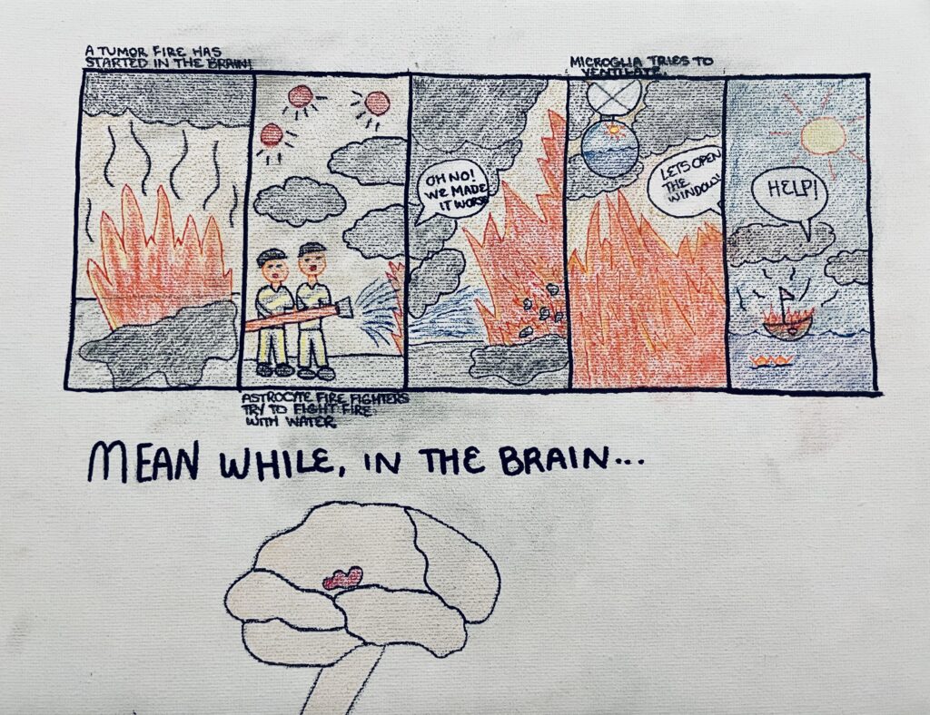
Despite a large amount of research into the cause of glioblastomas, not much is known about how they develop and why. Based on a trial which was published in 2018, the cancer cells which form glioblastomas originally come from the subventricular zone on the brain where mutated and cancerous neural stem cells are developed (Lee et al., 2018). Scientists and doctors alike, however, are still trying to figure out why this happens. What I would like to focus on, however, is the body’s response to this type of tumor once it has already begun to grow. In the scientific review Injury programs shape glioblastomas, the destruction caused by a tumor, in this case a glioblastoma, is compared to any other brain injury that may occur. The brain’s way of healing is the same (Brooks et al., 2022). This process involves both astrocytes and microglia. The purpose of astrocytes in the brain is to support neurons. These cells do so by regulating chemicals in the extracellular space, removing excess neurotransmitters, and responding to cell damage (OpenStax, 2017, p. 515). When a tumor begins to eat away at healthy brain tissue, the damaged cells release molecules called damage-associated molecular patterns, or DAMPs for short. These DAMPs are detected by astrocytes and microglia, telling the glial cells to begin either fixing or destroying damaged cells (Brooks et al., 2020). In my comic strip, this is represented by the first frame. An oil fire first is ignited, causing black smoke to fill the area. In my analogy, the fire is representative of a glioblastoma which is beginning to grow and damage cells around it. Black smoke represents the DAMPs being released by the dying cells. In the next frame of my comic, smoke alarms are sounded and a fire team attempts to fight the fire with water. This fire team represent astrocytes which are attempting to bring chemicals and nutrition to the damaged cells to help them recover. Just like how you would not want to fight an oil fire with water, you do not want to give a glioblastoma extra nutrients. When the astrocyte provides nutrients to the cells surrounding the tumor, it allows the tumor to grow just as it would do to a normal injured cell (Brooks et al., 2020). The third frame of my comic strip shows what happens when the astrocyte fire team puts water on the fire: the fire grows.
The second glial cell which I am focusing on for my project is microglia. The role of this cell in the brain is to remove damaged cells. Similar to macrophages in the rest of the body, microglia destroy and digest the damaged cells so new ones can take their place (OpenStax, 2017, p. 516). This process is represented in my comic in the fourth frame. The microglial fire team decides to open the window to rid the space of smoke, representing microglia eating DAMPs and dying cells. This too is an issue, however, as by opening the window they are providing the fire with oxygen and again causing it to grow. Microglial cells act similarly when confronted by tumor-damaged cells: they digest and try to eliminate the damaged cells. By doing this, they microglia are accidentally creating more space for the tumor to grow and spread to other regions of the brain (Brooks et al., 2022).
The work of both astrocytes and microglia, which in normal circumstances would help repair or replace damaged cells, effectively assists glioblastomas in their growth. Recent modeling of glioblastomas has shown an even ratio of both types of glial cells present in the tumor indicating their importance in tumor growth (Cornelison et al., 2022). This heterogeneity of cells present in glioblastomas is what makes them an aggressive and deadly type of cancer. Once diagnosed, the average survival time for a person with a glioblastoma who does not receive treatment is only 3 months. If a person is able to undergo tumor resection, radiation, and chemotherapy their survival increases to 15-16 months (McKinnon et al., 2021). Research continues on how to treat this disease that is seemingly supported by the body itself, however no cure has been found.
References
Betts, J.G., Desaix, P., Johnson, E., Johnson, J.E., Korol, O., Kruse, D., Poe, B., Wise, J.A.,
Womble, M., Young, K.A. (2017). Anatomy and Physiology. (515-516). OpenStax.
Brooks, L.J., Ragdale, H.S., Hill, C.S., Clements, M., Parrinello, S. (2022, November). Injury
programs shape glioblastoma. Trends in Neurosciences. 45 (11), 865-876. https://www.sciencedirect.com/journal/trends-in-neurosciences
Cornelison, R.C., Yuan, J.X., Tate, K.M., Petrosky, A., Beeghly, G.F., Bloomfield, M.
Schwager, S.C., Berr, A.L., Stine, C.A., Cimini, D., Bafakih, F.F., Mandell, J.W., Purow, B.W., Horton, B.J., Munson, J.M. (2022). A patient-designed tissue-engineered model of the infiltrative glioblastoma microenvironment. NPJ: Precision Oncology. 6 (54). https://doi.org//10.1038/s41698-022-00290-8
Lee, J.H., Lee, J.E., Kahng, J.Y., Kim, S.H., Park, J.S., Yoon, S.J., Um, J., Kim, W.K., Lee, J.,
Park, J., Kim, E.H., Lee, J., Lee, J., Chung, W., Ju, Y.S., Park, S., Chang, J.H., Kang, S.,
Lee, J.H. (2018, August 01). Human glioblastoma arises from subventricular zone cells
with low-level driver mutations. Nature. 560, 243-247. https://doi.org/10.1158/2159-8290.CD-RW2018-135
McKinnon, C., Nandhabalan, M., Murray, S.A., Plaha, P. (2021, July 14). Glioblastoma: clinical
presentation, diagnosis, and management. BMJ (clinical research ed.). 374, 1560. https://doi.org/10.1136/bmj.n1560
Glioblastoma is a brain cancer which develops in the subventricular zone of the brain. The brain’s response to repairing damage caused by this type of cancer is similar to that of any other type of brain injury and involves astrocytes and microglia. These two types of glial cells typically help to repair or replace damaged cells, however, in the case of glioblastoma, these cells cause the tumor to grow as if it were a typical injured cell.
Kelly’s visual depicts the process of a glioblastoma forming and is represented through the flames. The smoke represents the DAMPs that are released by the damaged cells. The water being put on the fire by the fire team represents astrocytes bringing nutrients to the glioblastoma. Because it is an oil fire, it should not be fought with water, just like glioblastomas should not be fed nutrients.