My project goes in more depth the stages of Mitosis. This is important because it is how our cells properly divide for every cell from skin cell to neuron cell within the body.
The cell cycle is comprised of three parts interphase, mitosis, and cytokinesis. According to chapter three of the “Openstax Anatomy and Physiology” textbook, “mitosis is the division of genetic material, during which the cell nucleus breaks down and two new, fully functional, nuclei are formed.” The major purpose of mitosis is to divide cells to form more cells to replace older cells. Interphase may range from a couple hours to many days. In contrast, the mitotic phase of the cell cycle usually takes between one and two hours. The four stages of mitosis include prophase, metaphase, anaphase, and telophase.
During prophase, chromatin condenses into visible chromosomes. These chromosomes become X-shaped. Courses.lumenlearning.com details that “sister chromatids begin to coil more tightly with the aid of condensing proteins.” The nucleolus disappears, and the nuclear envelope disintegrates. Also, centrosomes move to opposite sides of the cell and spindle fibers emerge from centrosomes. Spindle fibers, also known as mitotic spindles, are centrosomes with emerging microtubules.
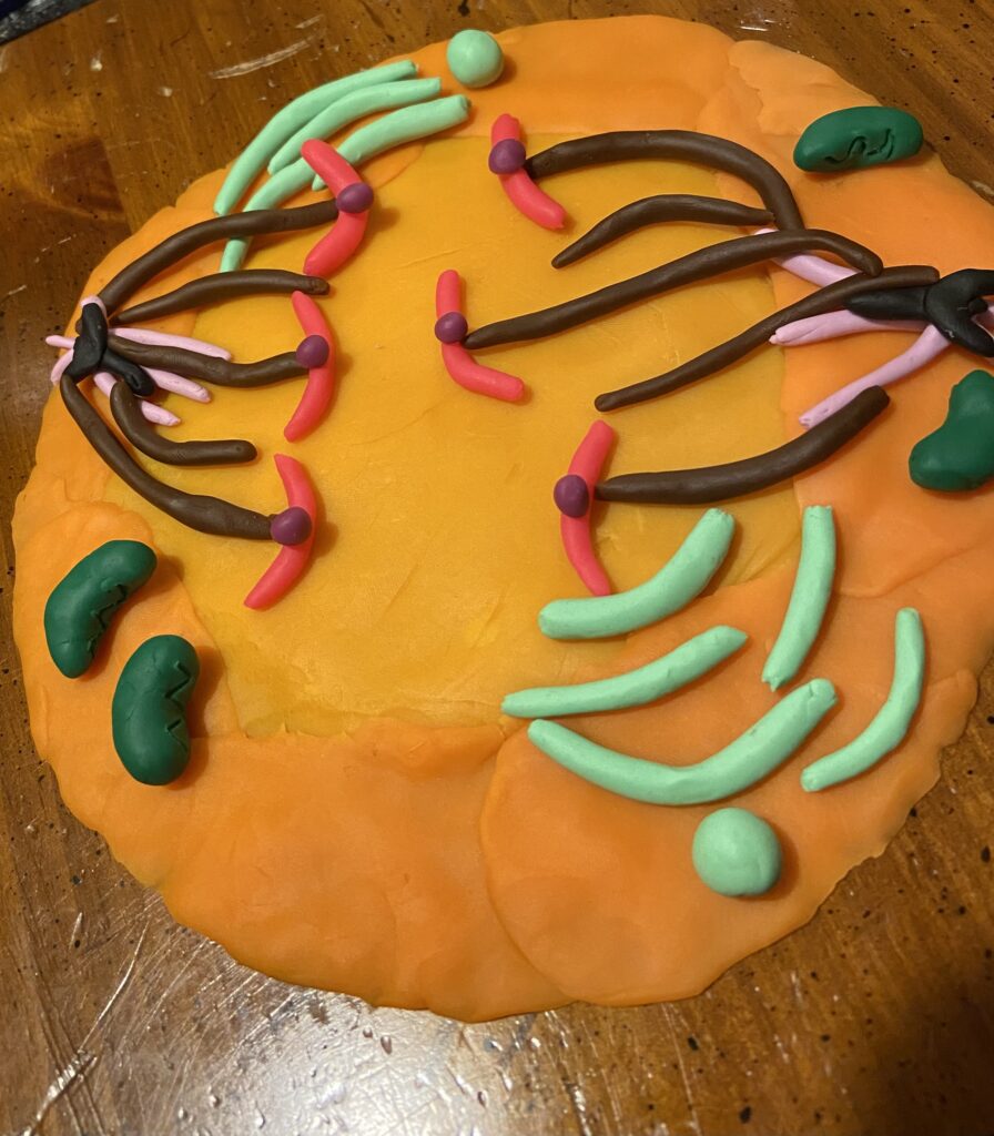
The next stage of mitosis is metaphase. Spindle fibers, attach to the kinetochore of the sister chromatids. The “Openstax Anatomy and Physiology” textbook, defines kinetochore as “a protein structure on the centromere that is the point of attachment between the mitotic spindle and the sister chromatids.” Khan Academy defines centromeres as “the regions of DNA where the sister chromatids are most tightly connected.” The metaphase plate appears during this stage which occurs when sister chromatids align in a straight line along the middle of the cell. According to Khan Academy, a spindle checkpoint “…helps ensure that the sister chromatids will split evenly between the two daughter cells when they separate in the next step. If a chromosome is not properly aligned or attached, the cell will halt division until the problem is fixed.” After the metaphase plate is formed and the spindle checkpoint is complete, the mitotic spindles begin to separate the sister chromatids by pulling them apart and bringing one chromatid from each pair to each end of the cell.
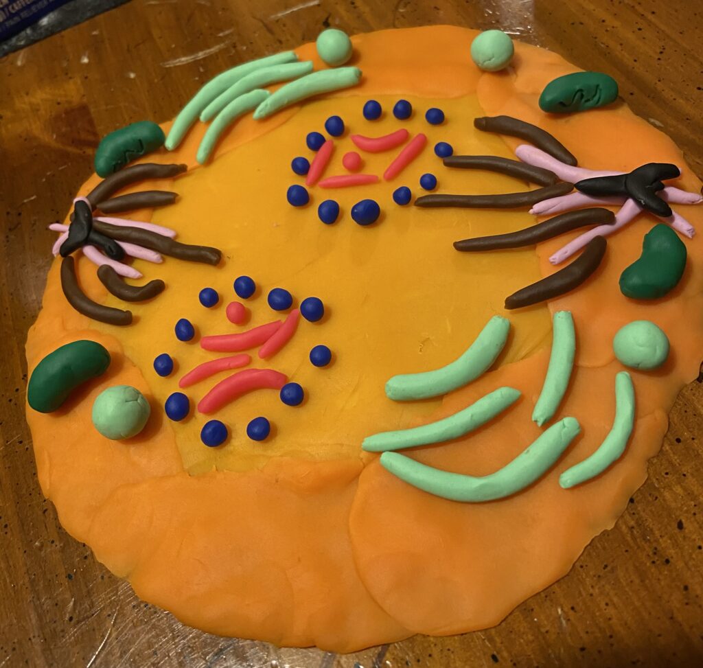
The third stage of mitosis is anaphase. Cohesion proteins that exist between sister chromatids break down to allow the centromeres to split in two. The sister chromatids transform into single chromosomes. The “Openstax Anatomy and Physiology” textbook explains that “each end of the cell receives one partner from each pair of sister chromatids, ensuring that the two new daughter cells will contain identical genetic material.” The chromosomes are then pulled to opposite ends of the cell toward the poles. Spindle fibers simultaneously shorten as the cell stretches and elongates forming an oval.
Telophase is the last phase of mitosis. Chromosomes arrive at the centrosomes that are located at opposite ends of the cell. When the chromosomes separate, mitotic spindle breaks apart and two new daughter nuclei form. The nuclear envelope appears in each nuclei and surround each set of chromosomes, unraveling the chromosomes which later becomes free flowing chromatin. Nucleoli reemerge in both nuclei, as well as DNA, organelles, membranes, and centrioles. Spindle fibers continue to push centrosomes apart until a cleavage furrow is created and cytokinesis begins.
References
Betts, J. G., Desaix, P., Johnson, E., Johnson, J. E., Korol, O., Kruse, D., Poe, B., Wise, J., Womble, M. D., & Young, K. A. (2017). 3.5 Cell Growth and Division. In Anatomy & Physiology (pp. 115–120). essay, OpenStax College, Rice University.
Khan Academy. (n.d.). Phases of mitosis | mitosis | biology (article). Khan Academy. Retrieved November 23, 2021, from https://www.khanacademy.org/science/ap-biology/cell-communication-and-cell-cycle/cell-cycle/a/phases-of-mitosis.
Learning, L. (n.d.). Mitosis. Biology for majors I. Retrieved November 23, 2021, from https://courses.lumenlearning.com/wm-biology1/chapter/reading-mitosis/.

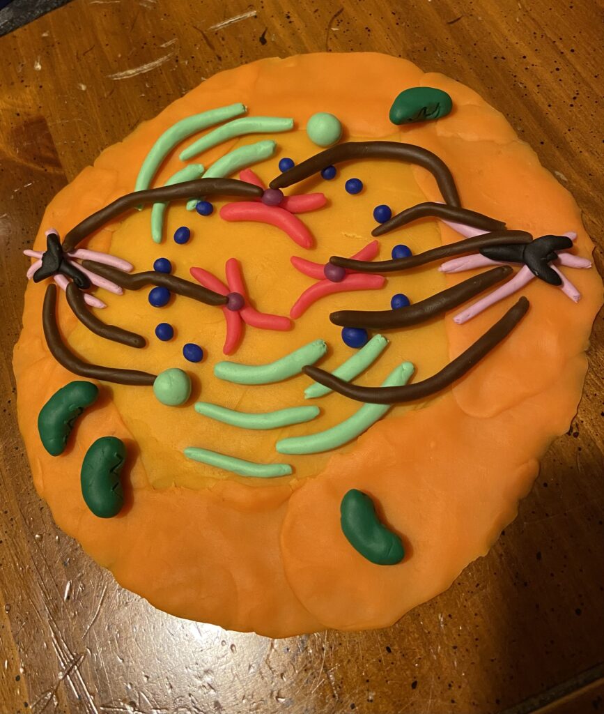
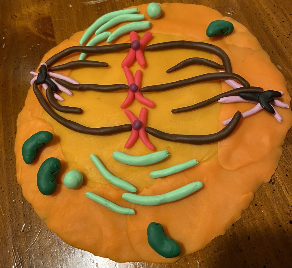
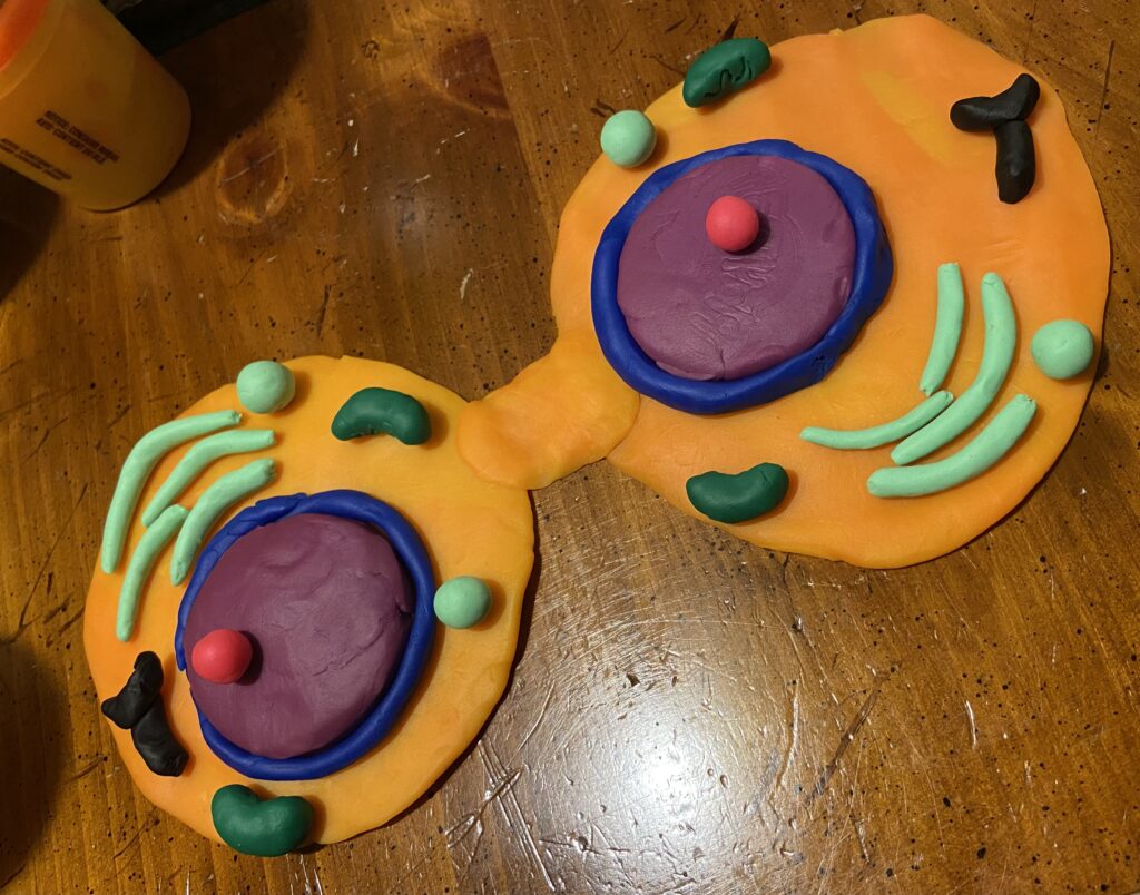
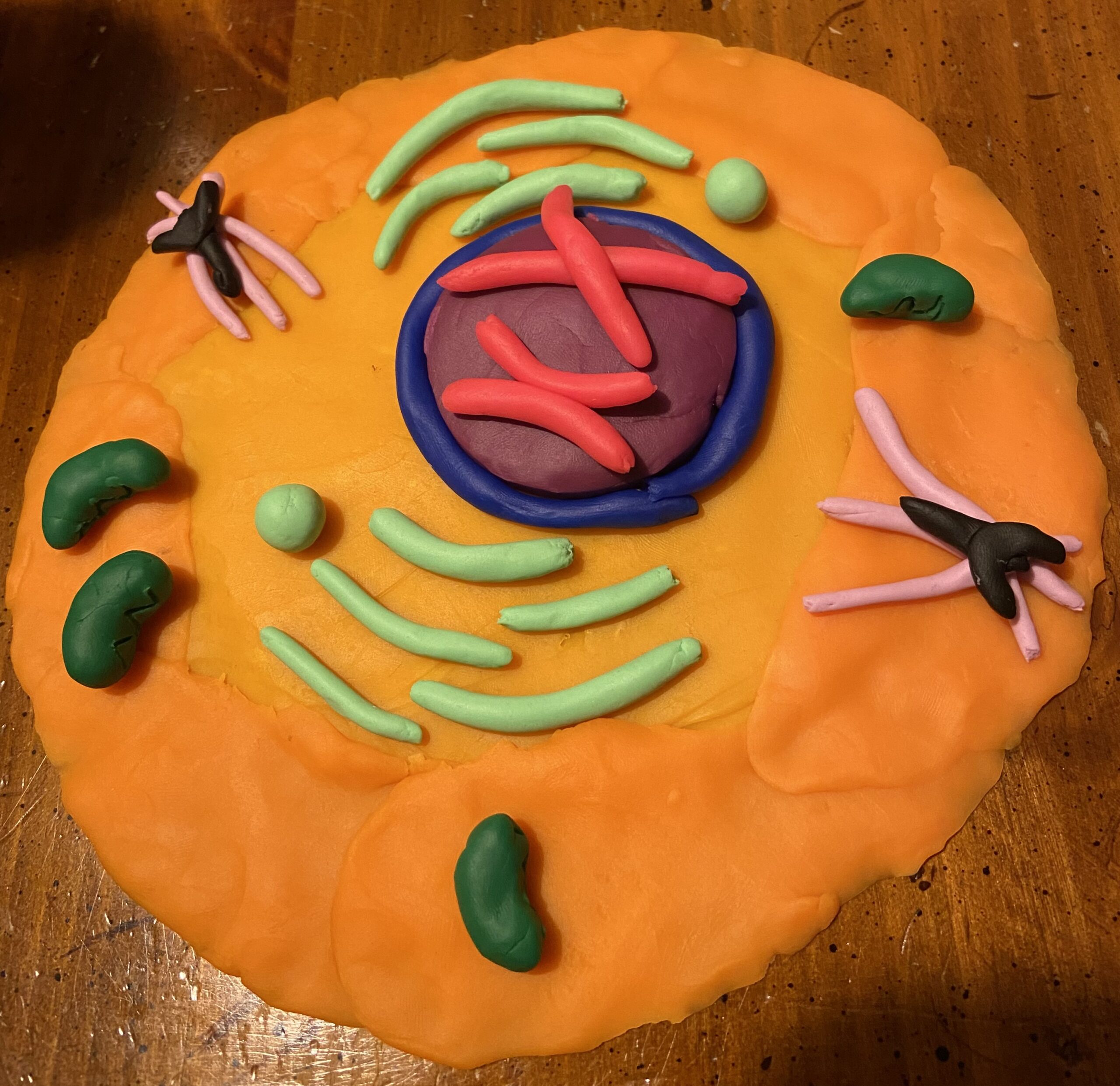
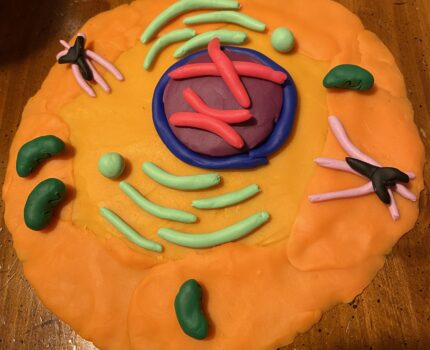
This project uses clay models in a series of six images to illustrate the different stages of the cell cycle, including interphase, all four stages of mitosis, and cytokinesis. The first image shows interphase, which is when the cell is at rest and not actively dividing. This is demonstrated by an intact blue and purple-colored nucleus and no active movement of the red-colored chromosomes or cell organelles. The second image shows prophase, where the blue nuclear envelope is shown to be breaking down into smaller pieces, and spindle fibers (shown here as long brown clay pieces) are forming to help move the chromosomes around. In the third image, which illustrates metaphase, the chromosomes are lined up in the center of the cell with the brown spindle fibers extending toward them outward from the cell poles. The fourth image demonstrates anaphase, where the chromosomes separate into sister chromatids and each one begins its movement toward an end of the cell. Telophase is shown in the fifth image, where new nuclear envelopes (again, small blue bits of clay) are shown forming around each set of chromatids. The sixth and final phase shows cytokinesis, where new nuclei have formed and the orange cell cytoplasm begins to pinch in the middle to form two new separate cells.