
STEAM Project
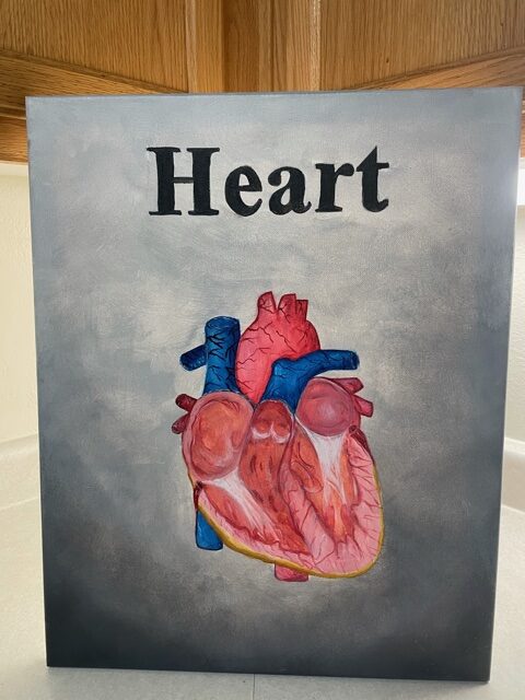
Structure and function the heart
I decided to do my STEAM project on the structure and function of the heart. The heart is described as a remarkable “pump” that propels blood into major vessels, the aorta and the pulmonary trunk. From these vessels the blood is distributed to the remainder the body (pg. 824). The human heart contracts approximately 108,000 times in one day more than 39 million times in one year. Each major pumping chamber of the heart ejects approximately 14,000 litters per contraction in a resting adult. The human heart is located within the thoracic cavity, between the lungs, the space is known as the mediastinum. The aorta and the pulmonary truck are attached to the superior surface of the heart, called the base (pg. 824). A typical hear is approximately the size of the fist 12cm (5 in) in length and 8cm (3.5 in) wide and 6 cm (2.5 in) in thickness. The female heart weights approximately 250-300 grams and the weight of a male heart is approximately 300-350 grams (pg.827). The human heart has four chambers, left and right each have one atrium and one ventricle.
The atria are the receiving chambers are small, thin-walled chambers that contribute little to propulsion of blood. The auricles are the appendages that increase atrial volume. The right atrium receives deoxygenated blood from body. The three veins empty into right atrium, The superior vena cava which returns blood from body regions above the diaphragm. The inferior vena cava, returns blood from body regions below the diaphragm. The coronary sinus returns blood from coronary veins. The left atrium receives oxygenated blood from lungs. Four pulmonary veins return blood from lungs. The discharging chambers make up most of the volume of heart. The right ventricle is the most of anterior surface. The left ventricle posteroinferior surface. The papillary muscles project into ventricular cavity. The anchor chordae tendineae are attached to heart valves, the thicker walls than atria, the actual pumps of heart, the right ventricle, the pumps blood into pulmonary trunk the left ventricle and the pumps blood into aorta (largest artery in body). The valves of the heart open and close in response to pressure changes. The two major types of valves are the atrioventricular valves located between atria and ventricles and the semilunar valves located between ventricles and major arteries. The two atrioventricular (AV) valves prevent back flow into atria when ventricles contract. The tricuspid valve (right AV valve) made up of three cusps and lies between right atria and ventricle. The mitral valve (left AV valve, bicuspid valve) made up of two cusps and lies between left atria and ventricle. The chordae tendineae anchor cusps of AV valves to papillary muscles that function to hold valve flaps in closed position and prevent flaps from everting back into atria.
The two semilunar (SL) valves prevent backflow from major arteries back into ventricles. It open and close in response to pressure changes. Each valve consists of three cusps that roughly resemble a half moon. The pulmonary semilunar valve are located between right ventricle and pulmonary trunk and the aortic semilunar valve are located between left ventricle and aorta.
The heart pumps equal volumes of blood throughout the pulmonary and systemic circuit. The pulmonary circuit is short and low-pressure circulation. The systemic circuit is long and high-friction circulation. The left ventricle walls are 3 times thicker than the right ventricle. The left ventricle pumps with more pressure.
Work Cited
Betts, J. Gordon. Anatomy & Physiology. OpenStax College, Rice University, 2013.
I like this idea of the project explanation for the heart process, thanks for sharing