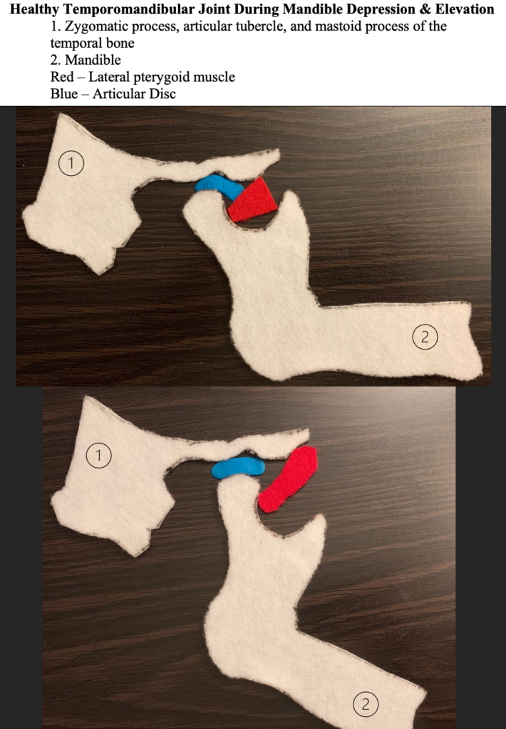
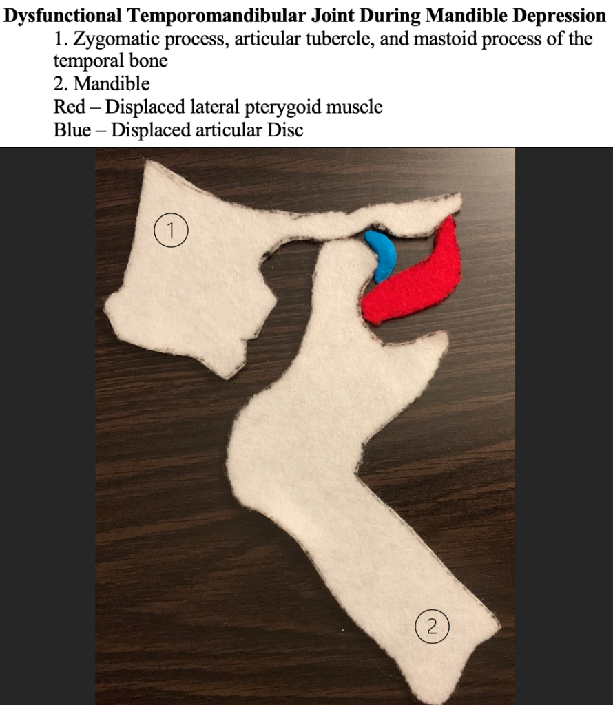
For this project, I chose to model temporomandibular joint dysfunction to meet the following objective: “relate the interaction of muscles and the skeletal system.” I chose this objective because temporomandibular joint dysfunction is largely caused by interrelated skeletal and muscular issues such as arthritis and extended muscle contractions. I used felt sheets to depict the temporomandibular joint which connects the mandible to the temporal bone and is cushioned by an articular disc. I made multiple models to show both healthy TMJ functioning and TMJ dysfunction which are distinguished by disc and muscle (dis)placement. The first two images following the written statement show healthy TMJ functioning during both mandible depression and mandible elevation. The last image shows TMJ dysfunction, specifically interrelated disc and muscle displacement, during mandible depression.
The temporomandibular joint is responsible for movements like mandibular depression, mandibular elevation, protraction, retraction, and lateral movement. A properly functioning temporomandibular joint’s articular disc is positioned between the condyle of the mandible and the mandibular fossa of the temporal bone. This position allows the lateral pterygoid muscle to pull the condyle of the mandible downwards while the articular disc glides forward, and the mouth opens. The lateral pterygoid also plays a role in stabilizing the articular disc, though this can become problematic as issues arise with the muscle (Lafrenière et al 1997).
Temporomandibular joint disorder, which causes a myriad of painful symptoms, can be caused by skeletal, muscular, or tissue damage; typically, damage or dysfunction of one temporomandibular structure exacerbates the dysfunction of another. Common causes include arthritis, disc or cartilage erosion, muscle fatigue, or jaw injury. For my project, I focused on the relationship between disc and muscle displacement which are usually caused by excessive strain or trauma. With TMJ disorders, instead of gliding forward with the hinge movement of the mandible, the articular disc often slips into an anterior position and may glide in and out of its proper position during mandibular depression. Articular disc displacement is sometimes caused by lateral pterygoid muscle spasms which exert forward traction on the disc (Finden et al 2007).
To elaborate on the symptoms, TMJ dysfunction can cause popping and clicking sounds, jaw pain, and limited range of motion including lockjaw. A recent study found that TMJ pain occurred significantly more in patients with disc displacement that caused jaw locking (Chantaracherd et al 2015). Because disc displacement removes the cushioning between the mandible and temporal bones, displacement can also damage the bones and cause or worsen osteoarthritis. Additionally, changes to the lateral pterygoid, including extended spasming or displacement, can cause the muscle to atrophy, further exacerbating symptoms. Overall, the interrelated nature of muscular and skeletal structures in the temporomandibular joint can cause worsening joint function throughout based on the damage or dysfunction of a single structure.
References
Chantaracherd, P., John, M. T., Hodges, J. S., & Schiffman, E. L. (2015). Temporomandibular joint disorders’ impact on pain, function, and disability. Retrieved from https://www.ncbi.nlm.nih.gov/pmc/articles/PMC4336155/
Finden, S., Enochs, W., & Rao, V. (2007, September 01). Pathologic Changes of the Lateral Pterygoid Muscle in Patients with Derangement of the Temporomandibular Joint Disk: Objective Measures at MR Imaging. Retrieved from http://www.ajnr.org/content/28/8/1537
Lafrenière, C. M., Lamontagne, M., & El-Sawy, R. (1997). The role of the lateral pterygoid muscles in TMJ disorders during static conditions. Retrieved from https://pubmed.ncbi.nlm.nih.gov/9586487/