
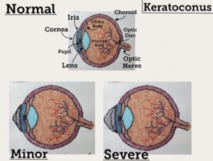
My STEAM project is on the corneal disease, keratoconus. My project shows three diagrams of the disease progression on an eye’s cornea in a cross stitch medium. The top one shows an eye in a normal, healthy state with various parts of the eye stated. The bottom left one shows an eye with minor keratoconus where the cornea is slightly bulging out in a small cone-like shape. Finally the bottom right one shows an eye with severe keratoconus where the cornea is now a large cone-like shape and is quite far from the pupil and iris. Both bottom diagrams has dotted lines in the cornea to show the difference between the affected cornea and a normal, healthy one.
Keratoconus is the thinning and scarring of the cornea. It tends to affects those between the ages of 10 to 25 years old the most, and takes about 10 or more years to become severe. There is no known cure for the disease, but there are treatments to slow down the progression such as specialized glassware and contact lenses for minor cases or corneal surgery if it gets severe. The cause of the disease is also unknown, but there are several theories.
The first theory is that your body starts producing too much of protease, an enzyme that breaks downs the collegen on the cornea, while producing too little of of the protease inhibitors. This leads to the body stripping down too much of the cornea’s collagen leading to to to thin and no longer support its shape. Another theory is that excessive abrasive actions, such as rubbing your eyes too much, or items, such as certain contact lenses, wears downs your cornea faster than your body can heal it back up. Another theory is that it could be hereditary, as according to some studies, as much as 10-15% of those affected by keratoconus has a family history of it. A final theory is that the disease is connected to other diseases/disorders such as allergies, asthma, and eczema. There appears to be link between these diseases and keratoconus as it is believed that the diseases create inflammatory reactions that affects the cells and collagen of the cornea.
When looking at the cellular level of a cornea, one would noticed that the cornea is made of both epithelial and connective tissues. The cornea is made up of unique stratified squamous cells that get oxygen from the air (the only body part to do so) and is the fastest healing body part, taking only 24-36 hours to heal back up. The cornea is also made of endothelial cells that contains collagen. This collagen is what helps the cornea to keep its small, round shape. When the cornea is afflicted with keratoconus, this collagen is then broken down slowly over a period of years causing the cornea to no longer to keep it small, round shape and instead bulge into a large cone-like shape. Since the cornea is the main part responsible for your vision on your eye, the distorted cornea has negative effects on your vision such as blurriness, double vision, sensitivity to light, loss of vision completely, and astigmatism.
Citations:
Ahuja, P., & Shetty, R. (2020). Relevance of IgE, allergy and eye rubbing in the pathogenesis and management of Keratoconus. Indian Journal of Ophthalmology, 68(10), 2067–2074. https://doi.org/10.4103/ijo.IJO_1191_19
Blackburn, B. J., Jenkins, M. W., Rollins, A. M., & Dupps, W. J. (2019). A review of structural and biomechanical changes in the cornea in aging, disease, and photochemical crosslinking. Frontiers in Bioengineering and Biotechnology, 7, 66. https://doi.org/10.3389/fbioe.2019.00066
Olivo-Payne, A., GodeFrooij, D., Chan, E., & Pedro-Aguilar, L. (2019). Optimal management of pediatric keratoconus: Challenges and solutions. Clinical Ophthalmology, 13(1), 1183–1191. https://doi.org/10.2147/OPTH.S183347
The topic of Adriana’s STEAM project was the corneal diseases Keratoconus. Adriana’s artwork was a series of cross-stitching that depicted what the cornea looks like at different stages of Keratoconus. The first picture was clearly labeled as to what each part of the eye was. All the cross-stitch pieces together effectively showed the major and minor changes that occur in the eyes of individuals with Keratoconus.
Adriana’s paper began by explaining what Keratoconus is, how it effects the eyes, and the difference between healthy eyes and eyes effected by Keratoconus. She then went over the various possible causes of the corneal disease. Lastly, Adriana explained the negative effects caused by Keratoconus.
I believe that her paper could have benefited from a closing statement, but overall, I think that Adriana did a good job conveying information about her subject. I did not know anything about Keratoconus previously, and I learned a lot from her paper and visual project. I thought it was very interesting how far the cornea protrudes away from the eye in individuals with Keratoconus. I enjoyed the cross-stitch visual as well, and I could tell that Adriana put a lot of work into this assignment.