
Paragonimus Westermani
Paragonimus westermani is a species of lung fluke acquired by a food-borne parasitic infection. P. westermani can cause a severe and fatal infection if left misdiagnosed and untreated. P. westermani is also known as the Japanese lung fluke or oriental lung fluke because infections are endemic in eastern Asia, but also common in Africa and South America.
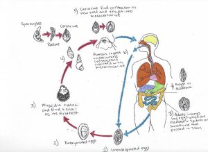
According to the CDC website, the parasite has a life cycle that includes two intermediate hosts and a final mammalian host. The eggs of P. westermani are excreted unembryonated in sputum, swallowed, or passed with stool. In an external environment, the eggs become embryonated, then miracidia enter its first host, a snail, by entering its soft tissue where the miracidia will go through three developmental stages; sporocysts, rediae, and cercariae. Once a cercariae leaves the snail and finds its second host, a crustacean, it will encyst and become a metacercariae. Humans become infected after ingesting undercooked or pickled crustaceans such as crab or crayfish that carry the metacercariae. The metacercariae hatch in the duodenum in the small intestine where they penetrate the intestinal wall and enter the peritoneum, then they continue to penetrate into the diaphragm and into the lungs, encapsulate themselves, and finally develop into mature adult flukes. Lung flukes can also penetrate into other organs such as the brain and liver to lay eggs, however, the eggs cannot complete their full life cycle because there is nowhere for the eggs to be released. Adult flukes range from 7.5 to 12mm by 4 to 6 mm in size and the time from infection to oviposition is 65 to 90 days. Humans can have an infection of P. westermani for up to 20 years. It can take one to two months or even longer for the infected person to experience any symptoms. Symptoms of infection from P. westermani can range from an acute cough, itching, diarrhea, abdominal pain, chest pain, discomfort, difficulty breathing, blood-tinged sputum and a low-grade fever. Lung flukes can eventually cause pulmonary abnormalities that mimic other conditions and diseases such as bronchitis, tuberculosis, and lung cancer. This causes P. westermani to be commonly misdiagnosed, especially in America where the infection is uncommon. Due to this reason, the infection is usually diagnosed by the identification of eggs found in sputum or stool samples, and antibody tests. In the article, “ Chest CT Features of North American Paragonimiasis’ Henry, et al., diagnostic tools such as chest x-rays and CT scans can clearly depict the damage caused by lung flukes such as pleural effusion; a build-up of excess fluid between the layers of the pleura outside of the lungs, inflammation, pleural thickening; leaving stripes thicker than 1mm, and pulmonary nodules; lesions and growth on the lungs made by tracks from the worm burrowing a tunnel.
According to the article “Pleural fluid characteristics of pleuropulmonary paragonimiasis masquerading as pleural tuberculosis’ by Hwang, et al., chest radiographs in patients with lung fluke infections commonly revealed low glucose levels, high LDH levels, cystic lesions, ground-glass opacities; lesions typically associated with lung cancer, and air-space consolidation from pulmonary edema caused by fluids such as pus, blood, or water build up, decreasing the rate of diffusion and affecting the lung’s ability to reach total lung capacity.
I’ve always enjoyed parasites and I loved both the visuals and the accompanying description of Paragonimus westermani.
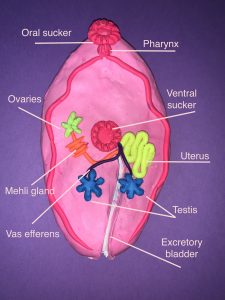
The most striking point of the clay replication is that Paragonimus westermani is hermaphroditic. A search reveled that all flukes (class Trematoda) along with class Cestodes are hermaphroditic EXCEPT for blood flukes, which are bisexual along with class Nematoda. From further reading, it appears that blood flukes and class Nematoda are also hermaphroditic but they do not self fertilize (really hard to find more information on bisexuality in parasites).
While most of the reproductive parts are similar in humans (ovaries, testis, vas efferens), I was unsure what the mehli gland. I discovered that it is a lubricated channel that directs the ova out of the fluke.
From the life cycle diagram and the description of the impact it had on humans, I was curious about the impact on the intermediate host of this parasite was but information was limited (most information is on how it impacts humans).
The most noticeable damage of P. westermani is to the lungs which was described in the tex though it mentions that it can and does travel throughout the body. It was mentioned that they can migrate to the brain. I found this paper which has CT scans of the brain of people infected with P. westermani. If the brain is infected, symptoms can include seizure, headache, vomiting, visual disturbance, and motor weakness.
While trematodes and other parasites may seem so rudimentary, they are quite complex especially to develop such an intricate reproduction and development pathway. Amazing creatures and amazing visuals plus description!