The purpose of this project is to show mitosis in a simple way to help educate people who have a hard time comprehending science or grasping concepts. It can be hard for some people to understand mitosis and also be able to remember it, this is why I chose to do this through a watercolor painting. Color has been shown to help create an “environment that fosters learning,” which is why it’s easier to learn mitosis when it isn’t black and white (Admin, 2019). I used a limited range of colors and added labels so when looking at my description of each step, you could reference what I’m talking about in my watercolor painting. I believe the colors allow you to see where everything is moving as well as what’s happening.

Description of Each Stage of Mitosis
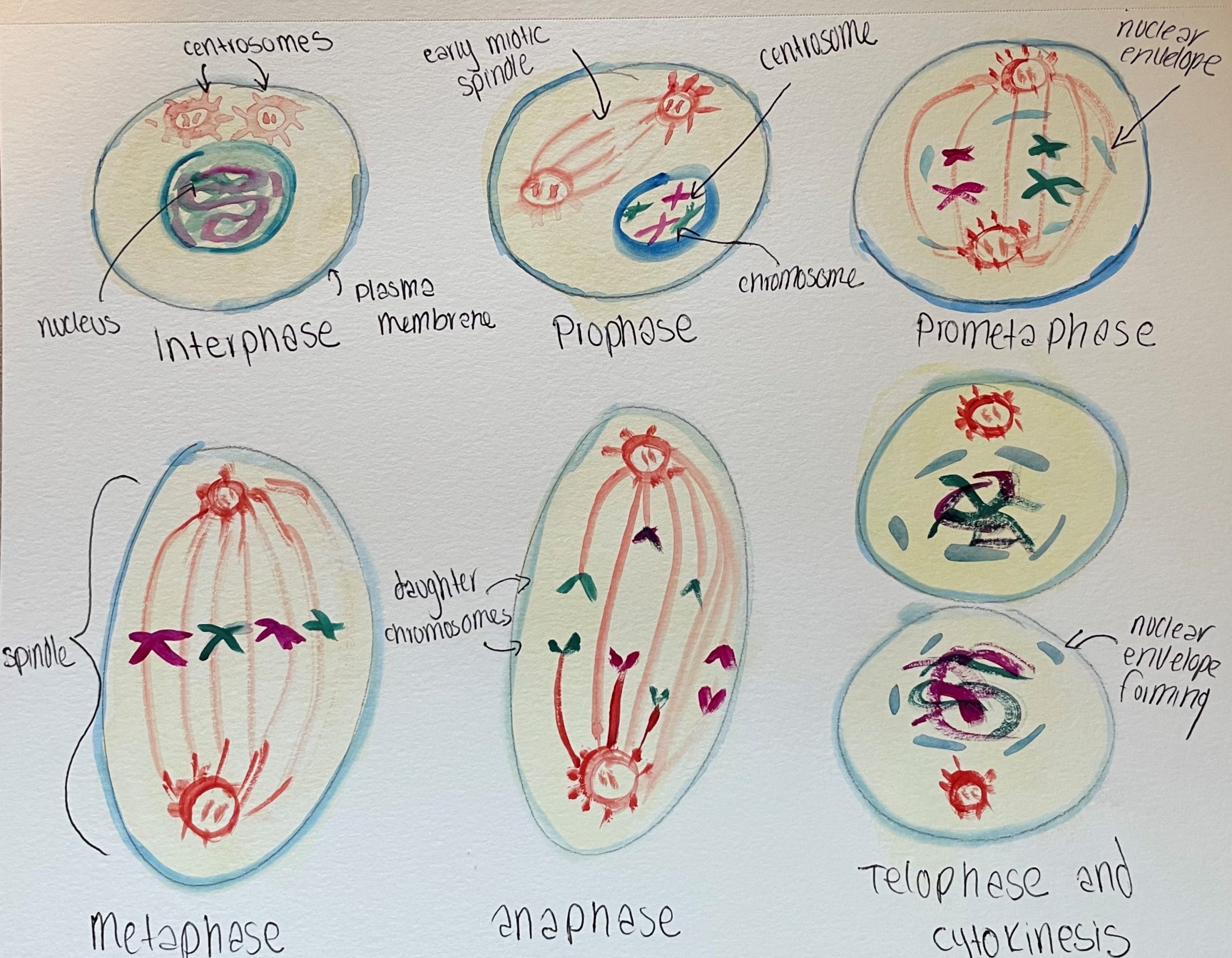
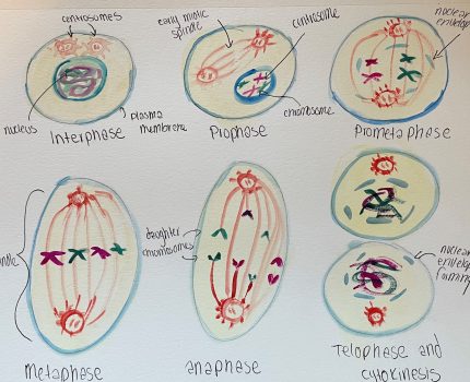
Interphase: The longest phase where the cell grows and prepares to divide. It makes a copy of its DNA which is found in the centrosomes (red). The nucleus is still present.
Prophase: The mitotic spindle (long red lines) begins to form between the centrosomes and the chromosomes begin to condense (purple and green). The nucleus is gone.
Prometaphase: The nuclear envelope (dark blue) begins to break down and the chromosomes fully condense. The centrosomes move to each side of the cell and the mitotic spindle is stretched between them.
Metaphase: The chromosomes line up in a straight line in the center of the cell, between the centrosomes.
Anaphase: The microtubules (which are the long red lines attached to the centrosomes) push the poles (centrosomes) apart and the other microtubules–called kinetochore microtubules–pull the chromosomes towards the poles.
Telophase and Cytokinesis: The chromosomes begin to condense and the spindle disappears. Now in two different cells, the nuclear membrane begins to form again and a nucleus appears. Cytokinesis will take place which will be different if it’s an animal or plant cell and it will form two cells.
Structures and Their Functions
Nucleus: controls and regulates the cell
Mitotic Spindle: divide chromosomes evenly in the original cell for the two resulting cells
Centrosomes: responsible for the organization of the microtubules
Plasma Membrane: provides protection and transportation for nutrients
Chromosome: Carries DNA
Nuclear Envelope: Keeps content of nucleus separate from the rest of the cell
Daughter Chromosomes: Result of mitosis and carries DNA
In the watercolor painting, you can see the different structures and how they play their role in mitosis. However, I would like to highlight two key points. In the painting, you can see the mitotic spindle, which is “assembled after [the] nuclear envelope breakdown” and how it is constantly working with the chromosomes (Rizzelli, 2020). This is because the mitotic spindle is responsible for splitting up the chromosomes between the daughter cells. However, the chromosomes aren’t accessible to the mitotic spindle until the chromosomes are broken out of the nucleus and mitosis can begin.
I would also like to point out that mitosis creates “two new daughter nuclei with equal genetic content” and creates two daughter cells (Moura and Carlos, 2019). This happens at the end of mitosis when “2 sets of chromosomes reach the spindle poles and the chromatin decondenses” before cytokinesis takes place (Fragkos and Valeria, 2017). The entire process of mitosis is necessary to create new cells to replace worn-out or dead cells and help with growth in the body.
I believe this project effectively communicates the steps of mitosis and leaves little room for confusion. I considered adding the description of each phase below each painting, but I decided a student wouldn’t feel as overwhelmed while learning mitosis if the process wasn’t crowded by text. I also wanted to remove the confusion about what each structure in mitosis does, so I added a description for the important structures in Mitosis, along with my description of each phase.
If I were to do this project again, I would try a different medium than watercolors. I think polymer clay would be a good idea since it would be a 3D structure. I also think it would be fun to focus on an idea relevant to mitosis such as cancer cells multiplying. However, I really liked gearing my project towards people who have a hard time understanding science.
Citations
Admin. “The Psychology of Color: How Do Colors Influence Learning?” Sh!Ft Disruptive Learning, 7 Mar. 2019, www.shiftelearning.com/blog/how-do-colors-influence-learning
Fragkos, Michalis, and Valeria Naim. “Rescue from replication stress during mitosis.” Cell cycle (Georgetown, Tex.) vol. 16,7 (2017): 613-633. doi:10.1080/15384101.2017.1288322 https://www.ncbi.nlm.nih.gov/pmc/articles/PMC5397263/
Moura, Margarida, and Carlos Conde. “Phosphatases in Mitosis: Roles and Regulation.” Biomolecules vol. 9,2 55. 7 Feb. 2019, doi:10.3390/biom9020055 https://www.ncbi.nlm.nih.gov/pmc/articles/PMC6406801/
Rizzelli, Francesca et al. “The crosstalk between microtubules, actin and membranes shapes cell division.” Open biology vol. 10,3 (2020): 190314. doi:10.1098/rsob.190314 https://www.ncbi.nlm.nih.gov/pmc/articles/PMC7125961/
Amazon https://www.npmjs.com/~amazon.us