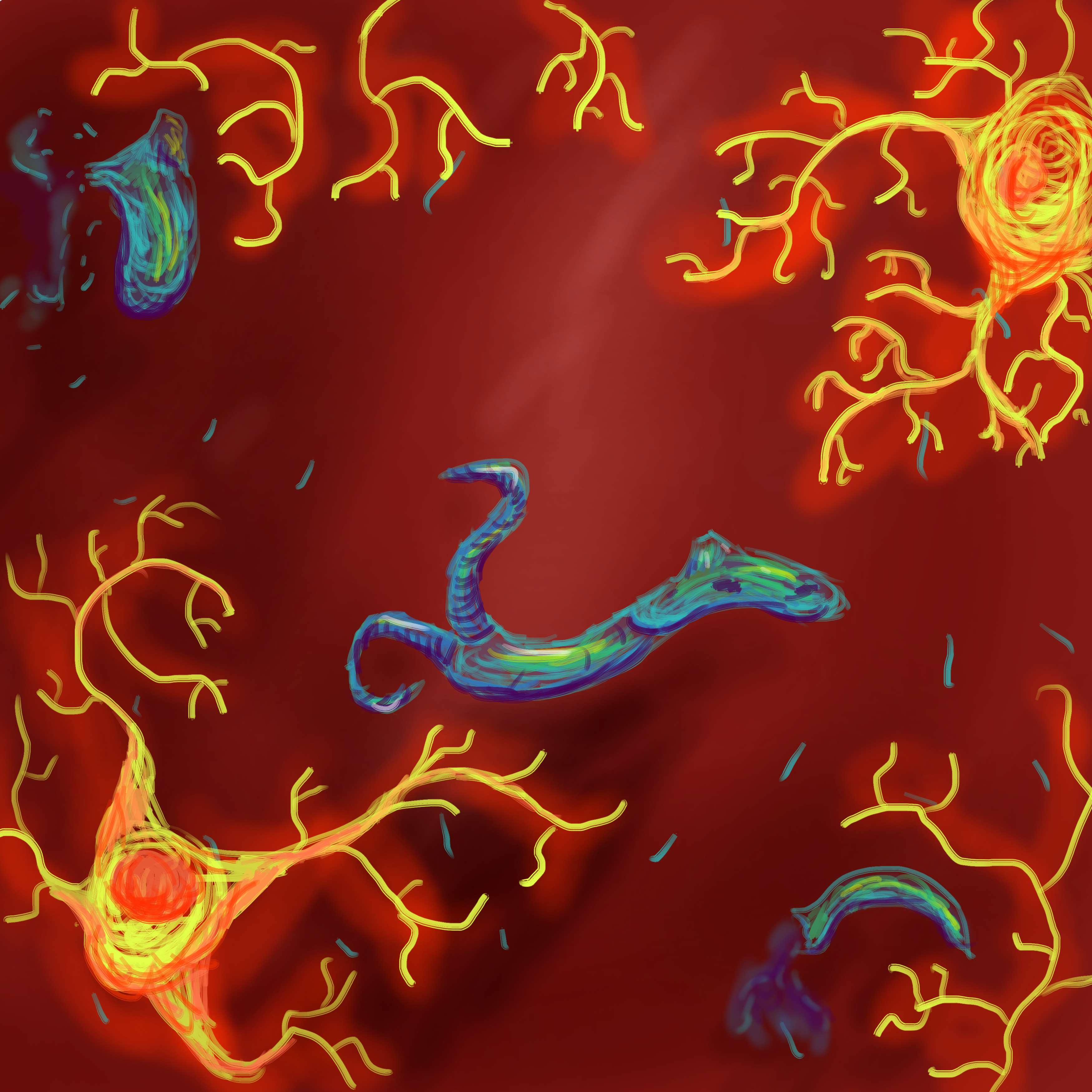
This relates to the objective of understanding the roles of nervous cells through the portrait of one of the strangest nervous cells.The image is of a set of microglial cells fighting an infection of Trichobilharzia Regenti in the brain. Although this is a more literal representation, it’s important to realize the immune function of microglial cells within the brains because it helps to better understand the bodily environment on the other side of the blood brain barrier. Besides the more misunderstood involvement in various aspects of homeostasis within the brain such as their relationship with amyloid plaques, the microglia protect the brain from what would otherwise be wholly unchallenged neurological invasion from parasites like T. Regenti as well as malaria.
T. Regenti infections in humans are purely accidental as they live in the CNS of birds and have a life stage in humans. Despite this, they can still cause damage to neural tissue in the brain while the infection lasts.
The role of the microglial cells helps to show the specific needs of the environment of the brain. Because of the lack of free circulating blood in the ventral cavities of the brain, this helps to protect it from most avenues of infection. In fact, free flowing blood in the ventral cavities would likely be impossible to live with practically due to the sheer volume of blood without vessel walls. The speeds needed to push blood effectively wouldn’t be reached unless the heart pumped with an exponentially more forceful beat than normal hearts. This lack of speed can also be attested by the slow production of CSF, as despite it’s motion being controlled by blood circulation, it doesn’t match the amount of equivalent blood flow in the cavity.
This lack of blood then exposes the brain to vulnerabilities to neurological pathogens due to the lack of blood carried immune cells. The microglial cells have then taken this role in order to allow for a more sterile environment. I chose this sort of image to begin with so I could highlight this role, as when I was looking into microglial cells, they were treated more like just another flavour of neuron. I think that they’re much more interesting and important than that, so I tried to draw them in a sort of dramatic way with the lighting. I don’t think that the image turned out quite how I’d like, but I think it’s ok.
I was originally going to have a swarm of microglial cells destroy the parasitic worm, but I decided to compromise so that the composition could include both the worm and microglial cells more prominently. I tried to make it less literal through the use of bold color to help liven the image from something more like a diagram and into a more fulfilled drawing. The part that best shows this is the orange glow around the dendrites, as I thought it helped to make them look more menacing. Having that can also help to connect with the idea of chemotaxis as well. Overall, I think it does a good job at trying to make people more appreciative of these cells.
Horak, P., Jones, M., Kolarova, L., Laddmann, H., Lichenbergova, L. (2011) Trichobilharzia
Regenti: Host Immune Response in the Pathogenesis of Neuroinfection in Mice. Experimental Parasitology, 128(4) 328-35. https://doi.org/10.1016/j.exppara.2011.04.006
.
Gonzalez, H., Elgueta, D., Montoy, A., Pacheco, R. (2014) Neuroimmune Regulation of
Microglial Activity Involved in Neuroinflammation and Neurodegenerative Diseases. Journal of Neuroimmunology, 274(1,2) 1-13. https://doi.org/10.1016/j.jneuroim.2014.07.012
Brinker, T., Klinge, P., Morrison, J., Stopa, E. A New Look at Cerebrospinal Fluid Circulation. Fluids and Barrier of the CNS, 11(10). https://fluidsbarrierscns.biomedcentral.com/articles/10.1186/2045-8118-11-10