
Many members of my family participate in hockey, wrestling, and martial arts. Plus, numerous other intensely competitive sports. I have witnessed practically all of my family members either in a cast or splint or through surgery for a fracture requiring pins or rods. Growing up, I understood that bones self-healed with time and gave humans their structure. That fracture may seem different depending on what you did when it occurred, such as jumping, hitting a wall, or slipping and landing on something hard. The so-called “boxer’s fracture” was typical in my household. Many people could develop what is sometimes referred to as the boxer’s fracture because it is one of the most common hand fractures that can occur.
Like me, most people discover their bones at a young age and that they are part of the skeletal system. Similar to how a house needs support beams, they are necessary. But the skeletal system serves a variety of purposes. It provides structural support, enables movement, safeguards internal organs, produces blood cells, and stores and releases minerals and fat, among other essential functions for the human body. Osseous tissue, a tough, dense connective tissue, comprises bones. In the areas of the skeleton where bones move (such as the joints and ribs), cartilage, a semi-rigid kind of connective tissue, provides a flexible and smooth surface for movement (Textbook Pg 214)Pg.
A fracture’s intricacy, location, and other characteristics are used to categorize it. Multiple terms may be used to describe some fractures. Transverse occurs directly over the bone’s long axis. Oblique, not at a 90-degree angle when it happens. Spiral A twisting motion causes bone fragments to be pulled apart. Comminuted There are several little bits between two main segments due to several breaks. Impacting one fragment usually results from compression driving the other piece into it. A fracture that only affects one side of the bone. Open or compound. A fracture resulting in at least one end of the shattered bone piercing the skin is likely to become infected. Closed, or simply a fracture in which the skin remains intact (Textbook Pg. 239).
When I was a kid and got cast out, I was told it would hold my bone in place so it didn’t move while it healed. The doctor just said to be patient, and it would fix itself. But how does bone repair itself when it is fractured? It starts Within 48 hours of the fracture, the endosteal chondrocytes and osteoblasts produce an outer callus of hyaline cartilage and bone, respectively, around the outside of the break (Textbook Pg. 240), while the endosteal chondrocytes secrete an internal callus (plural = calli) between the two ends of the shattered bone. As a result, the fracture is still stable. Osteoclasts resorb the dead bone over the next few weeks, while osteogenic cells become active, divide, and change into osteoblasts. During endochondral ossification, the trabecular bone replaces the calli’s cartilage. The internal and exterior calli eventually merge, compact bone replaces spongy bone at the fracture’s outer edges, and healing is finished (Fracture healing).
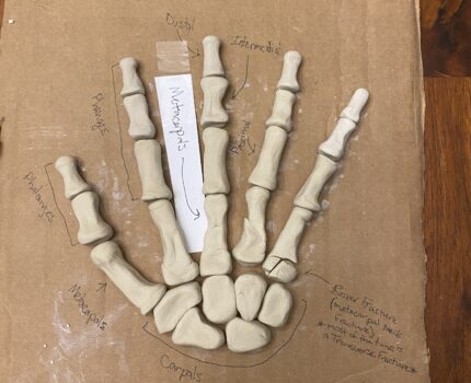
Another name for a fifth metacarpal neck fracture is a “boxer’s fracture.” Most of the time, it also falls under the Transverse type of fracture. According to the radiograph, the metacarpal head is dislocated, and soft tissue is inflamed (Bragg). All of my family members have experienced this fracture at some point. It is widely possible that it will happen to the general populace. A hockey player who punched another player received it. In another instance, a boxer’s hand was funny-landed during a martial arts competition, breaking it. However, given enough time and the right steps after seeing a doctor, fractures like this do not negatively impact people long-term.
References
Bragg, S. (2005, October). The Boxers’ Fracture. Redirecting. https://www-sciencedirect-com.uaf.idm.oclc.org/science/article/pii/S0099176705003260
Free textbooks online with no catch. OpenStax. (n.d.). https://openstax.org/details/books/anatomy-and-physiology
LaStayo, P. C., Winters, K. M., & Hardy, M. (2003, June). Fracture healing: Bone healing, fracture management, and current concepts related to the hand. Shibboleth authentication request. https://www-sciencedirect-com.uaf.idm.oclc.org/science/article/pii/S0894113003800030?via%3DihubSheen, J. R., Mabrouk, A., & Garla, V. V. (2023, April 8). Fracture healing overview – statpearls – NCBI bookshelf. National Library of Medicine. https://www.ncbi.nlm.nih.gov/books/NBK551678/
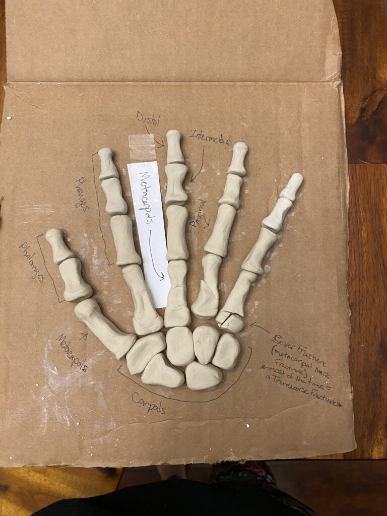
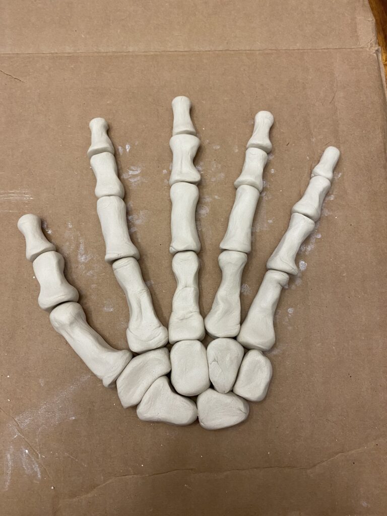
Kait’s project covers our course objectives of knowing the stages of bone development and repair and classification of fractures. Bone development first begins in the womb, continues throughout adolescence until epiphyseal plates close but osteoblasts and osteoclasts remain active throughout our entire life. Bone fractures are classified in a number of ways, intricacy and location are just a couple of ways fractures are classified. Common classifications of how the bone breaks include transverse, where a fracture occurs over the long axis of the bone, and oblique where the fracture occurs at an angle. Fractures can also be classified by if the skin barrier is broken by the skin, these fractures are known as open or compound fractures. Closed fractures are fractures where the bone doesn’t break the skin barrier. Kait focused on what is known as a Boxer’s Fracture. A Boxer’s Fracture is a break in the neck of the 5th metacarpal in the hand. A Boxer’s Fracture is typically classified as a transverse fracture and on a radiograph you can see the metacarpal head dislodged which will usually result in the surrounding soft tissue to be inflamed. A Boxer’s Fracture is typically casted to immobilize the bone. This immobilization allows for bone repair to begin. Within 48 hours, endosteal chondrocytes produce an outer callus of hyaline cartilage while osteoblasts will produce bone around the break. Endosteal chondrocytes will then secrete an internal callus in the bone gap, stabilizing the fracture. Osteoclasts will remove old bone while osteoblasts lay down new bone. Endochondral ossification begins, allowing for the trabecular bone to replace the calli’s cartilage. Finally the internal and external calli merge and spongy bone is replaced by compact bone on the outer edges. Kait’s art portion shows all of the bones of the hand.