
By: Eric Kelly
The course objective that I am going to cover in my STEAM project is “Know the stages of bone development and repair”. In order to fulfill this objective, I will start my project by briefly describing the stages of bone development of long bones before continuing on to discuss multiple methods of fracture repair.
Bone development:
There are two main ways that bone development can occur. Endochondral ossification will be the focus of this paragraph while the other way is called intramembranous ossification. The reason that this paragraph will primarily focus on endochondral ossification is that it is more common throughout the body and makes up most of the development in long and short bones (Setiawati and Rahardjo, 2018). According to Setiawati and Rahardjo the steps for endochondral bone formation begin with the mesenchymal cells grouping together in the shape of the bone and then changing into cartilage. After this process, the cartilage begins to grow and next capillaries turn the perichondrium into periosteum where osteoblasts can produce bone. From this point forward the cartilage continues to grow and get transformed into bone by osteoblasts (Setiawati and Rahardjo, 2018).
Bone Repair:
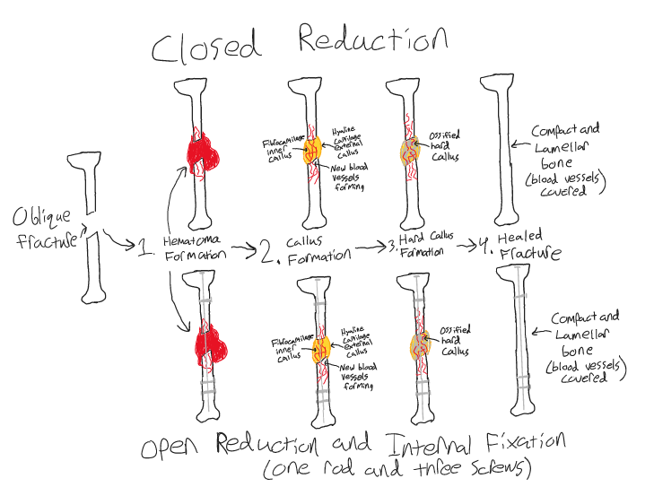
There are multiple different types of bone fractures, however, the one that will be discussed the most in this paper will be oblique fractures of long bones. There are multiple medical strategies to help with the repair of these fractures although the majority of the work is done by the body. The two strategies that will be discussed are closed reduction and open reduction and internal fixation (ORIF). According to the Cleveland Clinic “Oblique fractures occur when your bone is broken at an angle. The fracture is a straight line that’s angled across the width of your bone.” (Oblique Fracture: Symptoms, Causes & Treatment, n.d.). Due to the nature of oblique fractures the two ends of the bones can sometimes become offset which is where the two medical strategies come into play.
The first method mentioned is closed reduction. In this method, the bone is reset into a position where it can heal properly by a medical professional from outside of the body (Tibia/Fibula Fracture Open Reduction and Internal Fixation, n.d.). After this, the first step in bone repair will be the formation of a hematoma (Quinn, Kopp, and Vaughan, 2022). The formation of a hematoma clots the ruptured blood vessels and creates a temporary frame to begin initial healing (Sheen and Garla, 2022). The next step in repair is the formation of a fibrocartilaginous callus. In this step, chondrocytes begin to form a fibrocartilaginous structure in between the ends of the fracture with a sleeve of hyaline cartilage on the outside surrounding it while woven bone also forms near the periosteum (Sheen and Garla, 2022). After this, like in bone development, endochondral ossification begins to take place and the cartilaginous material begins to harden around the fracture into an ossified hard callus containing woven bone (Sheen and Garla, 2022) (Quinn, Kopp, and Vaughan, 2022). In the final stage of repair the bone will undergo remodeling to be the same as it was before the fracture. The callus in the center of the bone will be replaced by compact bone and the outer callus will be replaced by lamellar bone (Sheen and Garla, 2022) (Quinn, Kopp, and Vaughan, 2022).
The second method is open reduction and internal fixation (ORIF). The difference in between ORIF and closed reduction mainly involves the very beginning steps in which medical aid is used to help prepare a bone for natural healing. In this process, rather than a medical professional setting the fracture into a place where it can heal from outside of the body surgery is required. This method is usually only necessary for fractures that are more severe than others such as comminuted fractures but can still be used for fractures such as oblique. The first step in this method for an oblique fracture is going into surgery and the surgeon opening the fracture by making incisions in the skin. After this, the ends of the fracture can be placed in a position where they will be able to undergo natural healing. The next step involves internal fixation as either screws, plates, wires, or other types of metal can be attached to the ends of the fractures to hold them together. The fracture site can then be surgically stitched back together and the fracture will undergo the healing process (What Is ORIF Surgery?, 2021). The purpose of this process is to hold the fracture in a place where it will then be able to undergo the same healing steps that a fracture normally undergoes.
References
Aiyer, A. (2023, January 28). Fracture Healing – Basic Science – Orthobullets. Fracture Healing – Basic Science – Orthobullets. Retrieved November 23, 2022, from https://www.orthobullets.com/basic-science/9009/fracture-healingOblique Fracture: Symptoms, Causes & Treatment. (n.d.). Cleveland Clinic. Retrieved November 22, 2022, from https://my.clevelandclinic.org/health/diseases/22185-oblique-fracture
Quinn, C., Kopp, A., & Vaughan, T. J. (2022). A coupled computational framework for bone fracture healing and long-term remodelling: Investigating the role of internal fixation on bone fractures. International journal for numerical methods in biomedical engineering, 38(7), e3609. https://doi.org/10.1002/cnm.3609
Setiawati, R., & Rahardjo, P. (2018). Bone Development and Growth. In (Ed.), Osteogenesis and Bone Regeneration. IntechOpen. https://doi.org/10.5772/intechopen.82452
Sheen, J. R., & Garla, V. V. (2022, May 8). Fracture Healing Overview. Fracture Healing Overview – StatPearls – NCBI Bookshelf. Retrieved November 22, 2022, from https://www.ncbi.nlm.nih.gov/books/NBK551678/#_ncbi_dlg_citbx_NBK551678
Tibia/Fibula Fracture Open Reduction and Internal Fixation. (n.d.). Tibia/Fibula Fracture Open Reduction and Internal Fixation – Health Encyclopedia – University of Rochester Medical Center. Retrieved November 22, 2022, from https://www.urmc.rochester.edu/encyclopedia/content.aspx?contenttypeid=135&contentid=379
What Is ORIF Surgery? (2021, May 20). WebMD. Retrieved November 22, 2022, from https://www.webmd.com/a-to-z-guides/what-is-orif-surgery
Eric,
I learned so much about bone development and repair from your paper and illustration. In your paper, you discussed how the long bones in the human body work through these two processes. There are two main ways that bones develop, and these are endochondral ossification (more common) and intramembranous ossification (less common). Two of the most common bone repair methods are closed reduction and open reduction and internal fixation. In closed reduction, the bone that is broken is “set” back into place. This is normally done by a medical professional. This process does not happen by itself. The hope is that the bone will heal faster with this method. The first step in this process takes place when hematoma forms. Then a fibrocartilage callus forms. This material hardens later on. Finally, bone remodeling takes place. In open reduction and internal fixation, the bone is not “set” into place. A medical professional usually just lets the bone heal on its own after surgery. After going under the knife, the same bone repair process happens as the closed reduction method. The human body’s repair system is absolutely incredible. I like how you drew out both of these processes and added color to make each step clear. It is interesting how the final two outcomes of the bone look different, even though the recovery steps are the same. I recently went to the doctor and they were able to look at my bones through an X-Ray machine. The doctor concluded that I did not have an oblique fracture. Great work on your STEAM project!