
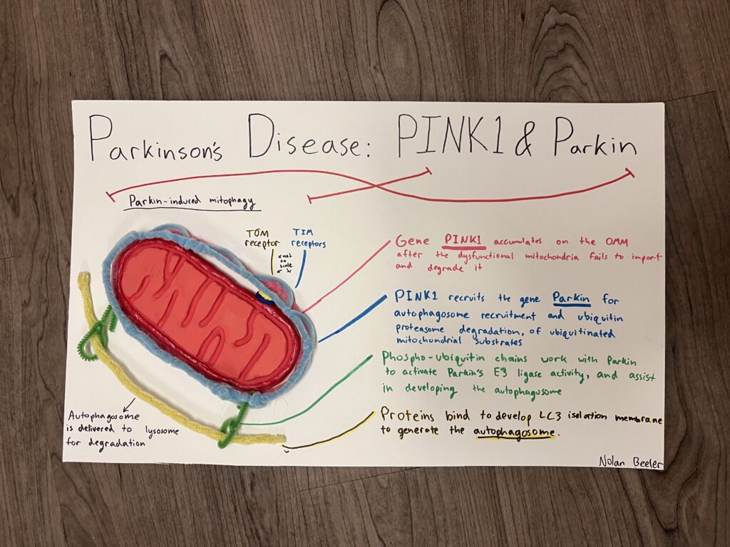
This project is a representation of the mutation of mitochondria in Parkinson’s disease patients. Parkinson’s disease (PD) is a degenerative nerve disease of the central nervous system. I chose this project as I was interested in different organelle functions and my grandma has parkinsons. Most PD sufferers develop symptoms past the age of 60, and the symptoms progressively get worse. These include stiffness, muscle tremors, slow movements, and balance issues among other things. It is still unclear what the exact cause of the disease is, and there is no cure. There are treatments such as deep brain stimulations, medications, and physical therapy that increase quality of life with the disease. PD is not fatal in itself, but as the symptoms progress over time, they lead to complications and issues with bodily function that often lead to death. Researchers have investigated what causes the death of nerve cells, and it’s possible that mitochondrial dysfunction leads to the death of these cells, as they cannot function without the cell powerhouse that is mitochondria. There have been multiple gene mutations found in both sporadic and familial PD patients. Most research and testing has been on patients with familial PD. Many genes appear to lead to PD, such as SNCA, LRRK2, but my project focused on the mutations of PTEN-induced putative kinase 1 (PINK1) and parkin (PRKN), and their role in mitochondria mitophagy. The model I created shows how a dysfunctional mitochondria, such as a mitochondria losing membrane potential, cannot process PINK1 and is degraded. My model is a clay mitochondria with pipe-cleaner chemicals that show the process of mitochondrial degradation in a dysfunctional mitochondria. In a healthy mitochondria, PINK1 is imported through the TOM and TIM complexes to be processed and produce peptidase. Once processed, the PINK1 is cleaved by the PARL protease, and then degraded by the proteasome. The PINK1 and Parkin genes usually only work together for mitochondrial quality control. In a dysfunctional mitochondria, like the one I modeled, the PINK1 is inhibited from being processed through the TIM complex, and gets stabilized on the surface of the OMM (outer mitochondrial membrane). PINK1 accumulates on the surface all around the mitochondria, resulting in phosphorylation of ubiquitin and recruiting of Parkin. The ubiquitin chains are recognized by autophagy adaptors. The autophagosome then transports the damaged mitochondria to the lysosome for degradation. The loss of these many mitochondria seem to lead to the impairment and death of neurons, resulting in less dopamine production. Researchers have looked upstream along the chemical pathway to determine if the regulators of PINK1 and Parkin have to do with their mutations. Kinases such as AKT have shown that with age, α-synuclein oligomers increase parkin auto-ubiquitination and lead to increased degradation. An approach for a therapeutic fix is mitochondrial enhancers that improve the function of mitochondria through transcription, translation, and import of nuclear-encoded components, and expression of genes, which showed positive results in lab rat experiments. Overall, there is still much research to be done on the role PINK1 and Parkin have is mitochondrial mitophagy, but scientists have gotten quite far in identifying causes of PD.
References
Borsche M, Pereira SL, Klein C, Grünewald A. Mitochondria and Parkinson’s Disease: Clinical, Molecular, and Translational Aspects. J Parkinsons Dis. 2021;11(1):45-60. doi: 10.3233/JPD-201981. PMID: 33074190; PMCID: PMC7990451.
Prasuhn J, Davis RL, Kumar KR. Targeting Mitochondrial Impairment in Parkinson’s Disease: Challenges and Opportunities. Front Cell Dev Biol. 2021 Jan 5;8:615461. doi: 10.3389/fcell.2020.615461. PMID: 33469539; PMCID: PMC7813753.
Alicia M. Pickrell, Richard J. Youle, The Roles of PINK1, Parkin, and Mitochondrial Fidelity in Parkinson’s Disease, Neuron, Volume 85, Issue 2, 2015, Pages 257-273, ISSN 0896-6273, https://doi.org/10.1016/j.neuron.2014.12.007.
Park JS, Davis RL, Sue CM. Mitochondrial Dysfunction in Parkinson’s Disease: New Mechanistic Insights and Therapeutic Perspectives. Curr Neurol Neurosci Rep. 2018 Apr 3;18(5):21. doi: 10.1007/s11910-018-0829-3. PMID: 29616350; PMCID: PMC5882770.
Prasuhn Jannik, Davis Ryan L., Kumar Kishore R., Targeting Mitochondrial Impairment in Parkinson’s Disease: Challenges and Opportunities, Frontiers in Cell and Developmental Biology, VOLUME 8, 2021, https://www.frontiersin.org/articles/10.3389/fcell.2020.615461 10.3389/fcell.2020.615461, ISSN=2296-634X
Gonçalves FB, Morais VA. PINK1: A Bridge between Mitochondria and Parkinson’s Disease. Life (Basel). 2021 Apr 21;11(5):371. doi: 10.3390/life11050371. PMID: 33919398; PMCID: PMC8143285.
This Piece is a clay sculpture of a mutated and dysfunctional mitochondria, with pipe cleaner to represent the gene (PINK1), Parkin, as well as phospho-ubiquitin chains and the autophagosome. This depicts the process of Parkin induced Mitophagy/degradation of mitochondria.
In this instance, the dysfunctional mitochondria is unable to process the gene (PINK1), and instead of being processed normally is transferred to the outer mitochondrial membrane (OMM), or the red pipe cleaner. The PINK1 gathered on OMM recruits Parkin, which becomes the blue pipe cleaner, and is recognized by the phospho-ubiquitin chains, or green pipe cleaner. The chains then interact with Parkin to develop the autophagosome, which is a key vesicle that will engulf the mitochondria and transport it to lysosomes for degradation.