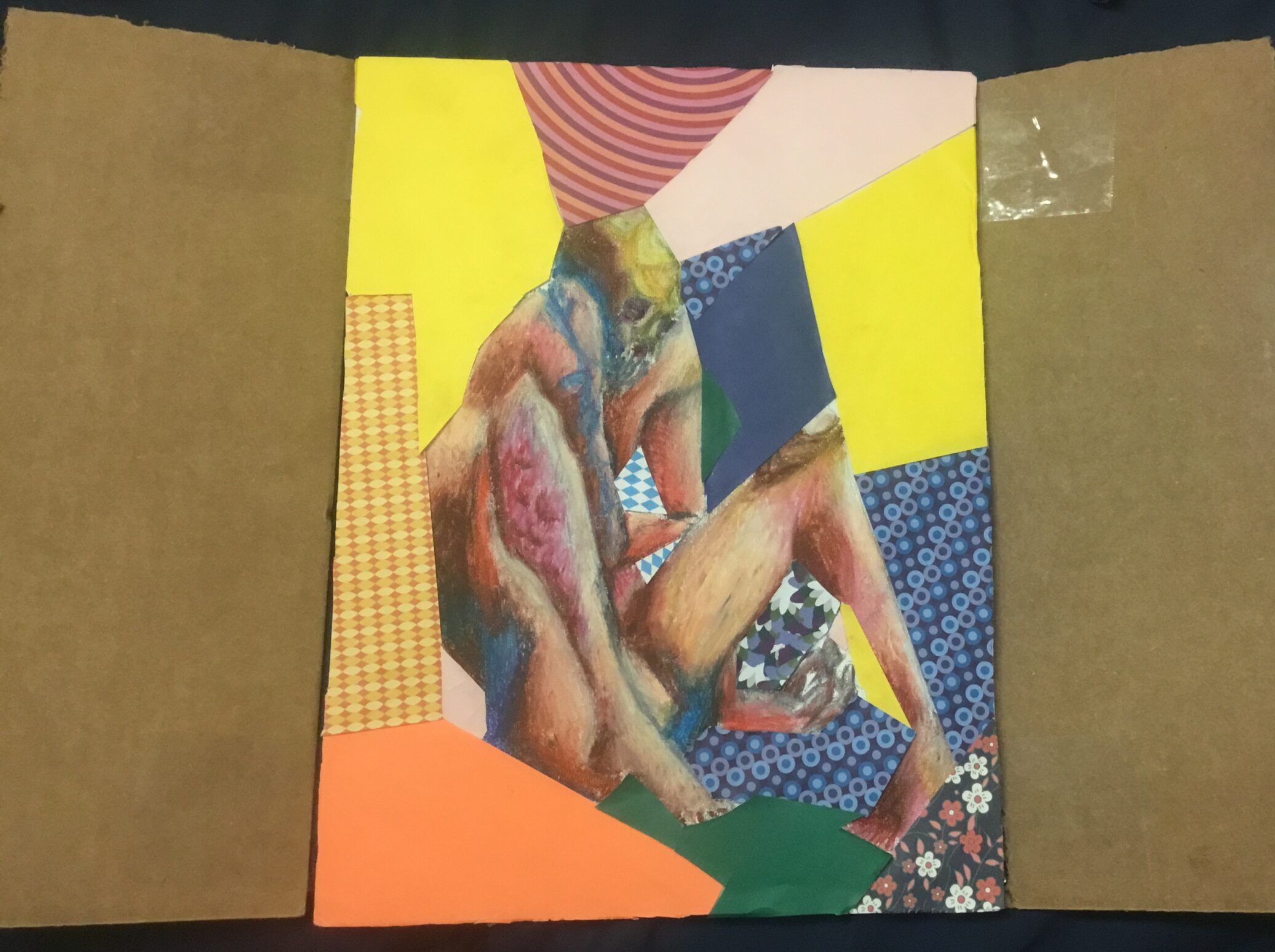
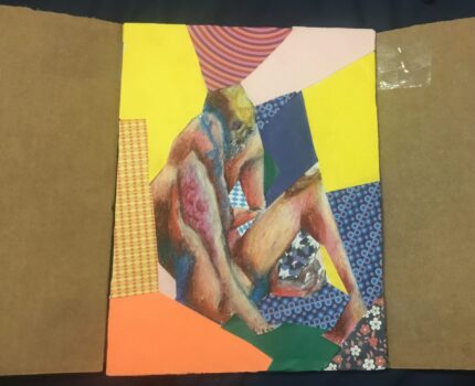
The piece is reminiscent of “skeletons in the closet”. As such, the outer layer depicts two opening closet doors. The image shows a corpse with its body parts in various stages of decomposition, surrounded by colorful papers. I used oil pastels, cardboard, and origami paper. With this image, I depicted different tissues in various states of decomposition with the intent to cover the objective “Describe the 4 major tissue types”, going into how the tissues change post mortem.
The four major tissue types are muscle, epithelial, connective, and nervous. The figure is drawn in an unstable pose to imply rigor mortis, caused by muscle tissue. As the body dies, it ceases to produce ATP, which prevents the muscles from relaxing as the cells can no longer release calcium. As decay continues, rigor mortis will end as the muscle cells decompose and break apart. As the cardiac muscle tissue of the veins no longer moves, the blood begins to pool to parts that are nearer to the ground, which is visible on the figure’s left hand and foot. In addition to blood pooling, marbling will occur which is on the right arm. This marbling occurs quickly after death and becomes permanent after around eight hours, but this pattern will become difficult to see as the skin and flesh darken. While I depicted the marbling to be blue to increase contrast from the skin, it is more common to appear as a deep purple or red. As blood cells decay, they stain the vein walls which helps expose the superficial veins during marbling. The epithelial tissue is shown in various states, with the figure’s left knee depicting skin slippage. During decomposition, the epithelial tissue dries out and becomes leathery. As the skin becomes dry and hard, it becomes easier to tear, and without the ability to create new cells, friction and environmental stress eventually cause the skin to tear and “slip off”. While nervous tissue isn’t explicitly shown in the image, the effects on this tissue are still visible. With no ability to send or produce signals, the body remains still. Under the surface, this tissue is softening. The gray and white matter of the brain become less distinguishable as the tissues mush together, liquifying while muscle tissue takes a detour and stiffens first. Bone is tissue that will end up lasting the longest in a recognizable form. This connective tissue dries out and becomes much more fragile post mortem. As the process of breaking down and rebuilding can no longer be performed, microfissures which would have once led the bone to become stronger, add together to crack and potentially shatter the bone. The skull of the figure is visible, using brown and yellow colors to imply dryness. Another connective tissue, adipose fat, will turn into adocipere. Otherwise known as “Grave Wax”. This wax forms predominantly in fatty areas such as the breast, buttox, and stomach. There are hints of its formation in the image with white speckling most noticeable on the thigh.
Yadav, A. B., Angadi, P. V., Kale, A. D., and Yadav, S. K. (2015). Histological assessment of cellular changes in postmortem gingival specimens for estimation of time since death. Journal of Forensic Odontostomatology, 33(1): 19–26. PMC5734814
Lee Goff, M. Early post-mortem changes and stages of decomposition in exposed cadavers. Exp Appl Acarol 49, 21–36 (2009). https://doi.org/10.1007/s10493-009-9284-9
The Australian Museum. (n.d.). Stages of decomposition.https://australian.museum/learn/science/stages-of-decomposition/
This is a really cool project, Clyde! I found the analogy of the closet doors to be really insightful., and the use of such bright colors was really enjoyable to study. I wasn’t aware of the marbling that would take place after rigor mortis sets in, but it makes sense given the lack of blood flow to the extremities. The drying of the bone was displayed in a really cool way, and I liked that you used colors such as yellow and brown to show this occurring. I also had never heard of the term “grave wax”, which you mentioned forms in fattier areas of the body, and after researching it more I learned that this is caused by the body fat being exposed to bacteria, mainly in warmer areas such as soil or water. As for the epithelial tissues, the way that you depicted the slippage in the figure’s left knee was also really interesting! I really enjoyed learning more about this topic from your project!