 Posted by

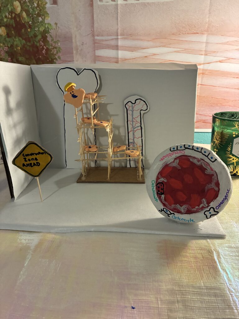
Bone tissue is made up of 5 different bone cells: osteogenic/stem cells, osteoblasts, osteocytes, osteoclasts, and bone-lining cells.
Osteogenic cells, also known as osteoprogenitor cells, are stem cells found in the bone marrow and periosteum that have the ability to differentiate into osteoblasts or bone lining cells. Osteogenic cells are a part of a broader group called skeletal stem cells (SSC), and are a type of SSC called mesenchymal stem cells (MSC) that have undergone differentiation and are committed to bone-forming (Matsushita & Ono, 2020). Skeletal Stem Cells are involved in producing not only bone, but also cartilage and fat cells. MSCs are also known as Bone Marrow SSCs due to their preferential localization near bone marrow sinusoids, or blood vessels (Matsushita & Ono, 2020).
Osteoblasts are cuboidal cells located along the surface of the bone, which are derived from mesenchymal stem cells. Osteoblasts are involved in bone formation and therefore tend to have larger numbers of Endoplasmic Reticulum and Golgi Bodies for higher rates of protein synthesis (Florencio-Silva et al., 2015). Mature osteoblasts have the ability to become osteocytes or bone-lining cells if they do not undergo apoptosis.
Bone-lining cells are flat cells that cover both internal (endosteal cells) and external (periosteal cells) bone surfaces. In these areas, neither bone resorption or formation takes place. It’s believed that the function of these cells includes maintaining the bone matrix by preventing interaction with osteoclasts so that bone resorption does not occur, as well as potential function in osteoclast differentiation (Florencio-Silva et al., 2015).
Osteocytes have the longest life span of all the bone cells, and are also the most abundant, making up between 90-95% of total bone cells (Florencio-Silva et al., 2015). Osteocytes are indirectly formed from MSCs during osteoblast differentiation, which takes place over 4 distinct stages (Florencio-Silva et al., 2015). During this differentiation process, the cytoplasm’s size decreases relative to the nucleus, and the number of organelles such as ER and Golgi body also decrease, reflected by lower levels of protein synthesis (Florencio-Silva et al., 2015). Osteocytes are found within the lacunae, and are believed to have several different functions as mature bone cells (Florencio-Silva et al., 2015), mainly including maintaining the bone matrix along with bone-lining cells as well as acting as a communication mediator for other types of cells during bone remodeling, as well as responding to mechanical stimuli as a stress sensor.
Osteoclasts are multinucleated cells involved in bone resorption. Mature osteoclasts are found on the bone surface, in resorption bays known as Howship lacunae. These cells are derived from a unique stem cell type known as hematopoietic stem cells (HSC), which are closely related to but not a type of SSC (Florencio-Silva et al., 2015), and which are found in the bone marrow before differentiation. HSCs are also involved in producing blood cells (Lee & Hong, 2020). Osteoclast activity is especially relevant in osteoporosis research.
Bone injuries can occur from many different causes and at different severities, and although bone has the potential to heal and reform through natural bone remodeling, this process may be much slower or more difficult for severe injuries or specific patient populations, such as older patients who have lower osteoblast activity (Domingues et al., 2015). Injuries requiring additional assistance for bone remodeling typically undergo the process of bone grafting, though certain difficulties have arisen with this method, including deficiency in donor availability, and prioritizing compatibility potential for proper regeneration (Su et al., 2021). New technology has allowed for new methods in the medical field to form and develop in recent years. Bone Tissue Engineering (BTE) is a new medical field that uses biomaterials, cells, and growth factors to synthetically boost bone remodeling. Stem Cell Therapy is a main part of BTE that consists of using stem cells, most notably Mesenchymal Stem Cells (MSC) for bone regeneration.
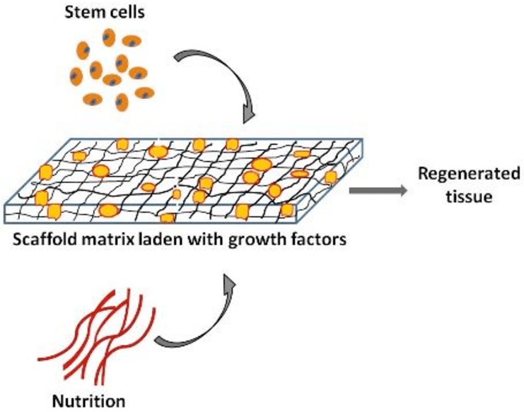
There are a few different methods that have been developed for stem cell therapy in bone regeneration. One method involves injecting stem cells, sometimes from a different area of the patient’s own body, into the necessary location; however, this is not as effective for larger-scale injuries (Su et al., 2021). Another method that has been in development in recent years is the construction of an engineered 3D printed scaffold made up of mesenchymal stem cells and additional growth factors that promote regeneration (Domingues et al., 2015). Scaffolding in bone tissue engineering is the implementation of a temporary, synthetic extracellular matrix (ECM) in situ meant to promote bone formation (Su et al., 2021) at the given site. There are two main methods in creating 3D printed scaffolds for stem cell therapy; one known as dECM (decellularized extracellular matrix) incorporation, where mesenchymal stromal cell-derived dECM is grown in vitro with the scaffold, causing the scaffold to homogenize with the dECM (Domingues et al., 2015). Another method, which is more commonly used, is known as dECM decoration, where dECM is deposited on the scaffold afterwards, causing it to bind only to the exterior of the scaffold (Domingues et al., 2015). To create a successful scaffold, certain factors are required. These factors include porosity and surface area ratio, as well as biocompatibility, and mainly whether or not the activity is matching the required clinical application, as well as if it can effectively promote cell adhesion, growth of blood vessels, and nerves (Su et al., 2021). In terms of shape, it has been found that squared scaffolds appear to promote the best MSC activity (Su et al., 2021). Overall, bone tissue engineering and stem cell therapy through the implementation of scaffolds, specifically those developed through 3D printing, shows lots of potential as an evolving method to combat the issues associated with serious bone injuries and the increasing need for stem cells in bone remodeling for treatment, though research is still underway.
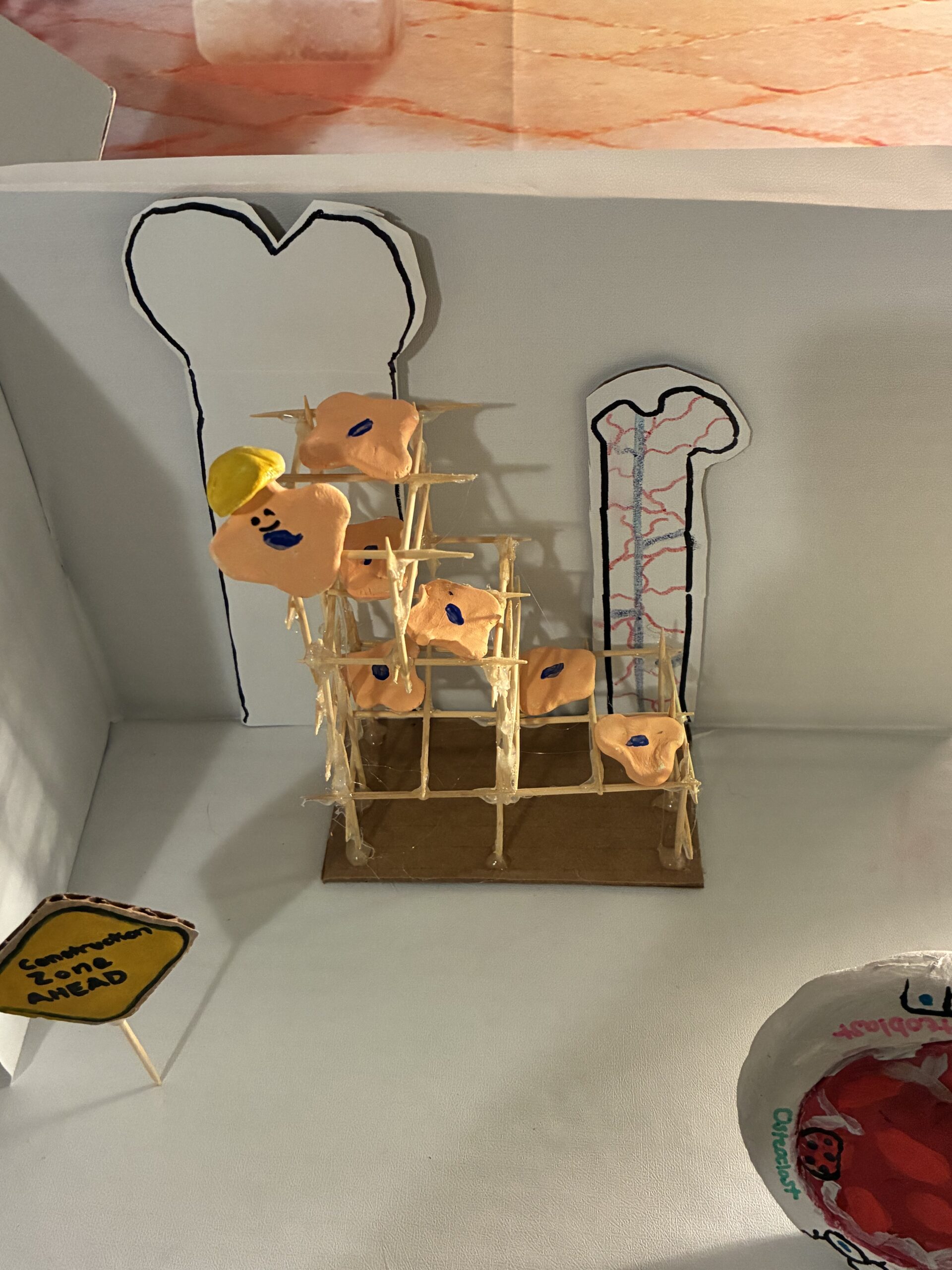
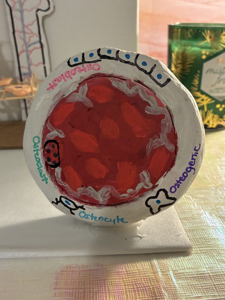
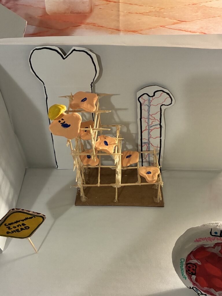
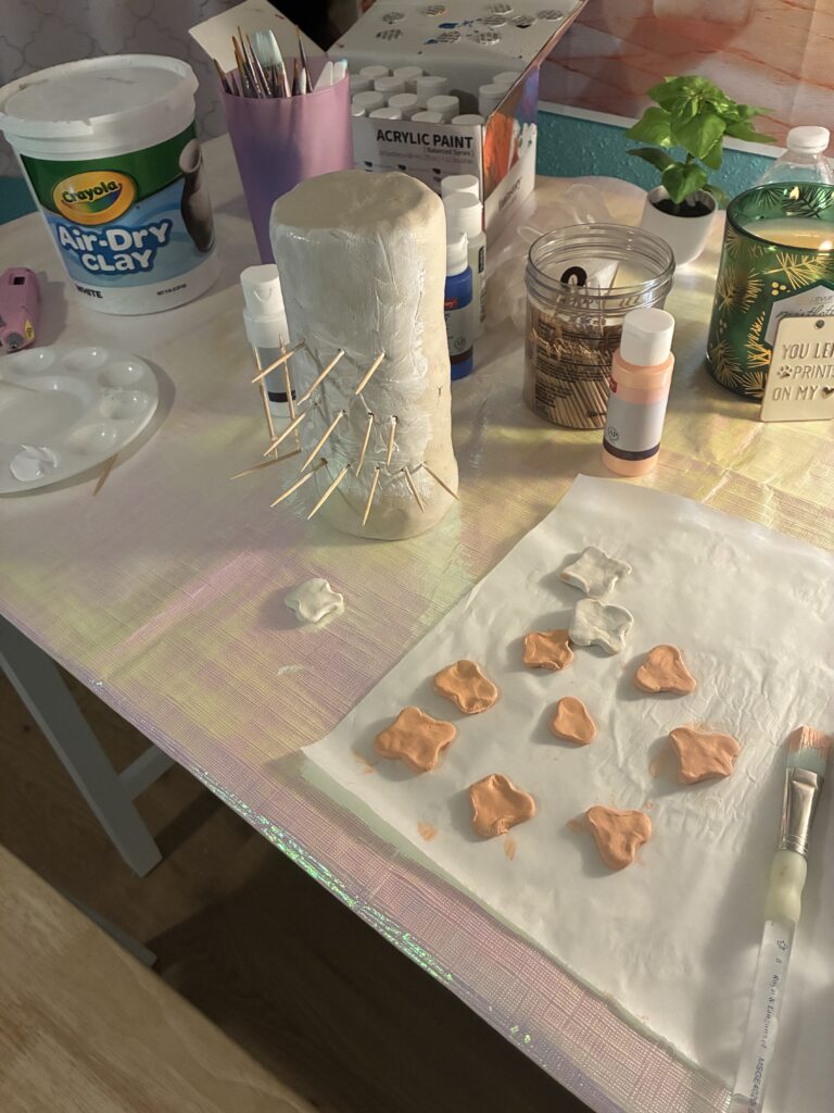
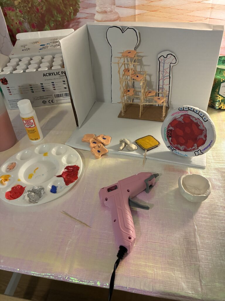
The project is meant to be an abstract 3D representation of the engineered scaffolds used for bone reconstruction, which are grids of biopolymers or matrix interwoven with stem cells to provide structure and support new bone growth. The scaffolds, similar to a microscopic version of those used in building construction, provide support along an injury site so that stem cells can work to generate new bone in the area. The project also includes a 3D diagram of the locations of 4 main bone cells covered in the focused objective: “Identify the 4 cells that comprise bone tissue.”
In Progress Photos:
References
Domingues, M. F., Carvalho, M. S., Sanjuan-Alberte, P., & Silva, J. C. (2025). Synthetic scaffolds functionalized with mesenchymal stem/stromal cells-derived extracellular matrix for bone tissue engineering: a review. RSC Advances, 15(38), 31812–31829. https://doi.org/10.1039/d5ra02106e
Florencio-Silva, R., Sasso, G. R. da S., Sasso-Cerri, E., Simões, M. J., & Cerri, P. S. (2015). Biology of Bone Tissue: Structure, Function, and Factors That Influence Bone Cells. BioMed Research International, 1–17. https://doi.org/10.1155/2015/421746
Lee, J. Y., & Hong, S.-H. (2019). Hematopoietic Stem Cells and Their Roles in Tissue Regeneration. International Journal of Stem Cells, 13(1). https://doi.org/10.15283/ijsc19127
Matsushita, Y., Ono, W., & Ono, N. (2020). Skeletal Stem Cells for Bone Development and Repair: Diversity Matters. Current Osteoporosis Reports, 18(3), 189–198. https://doi.org/10.1007/s11914-020-00572-9
Su, X., Wang, T., & Guo, S. (2021). Applications of 3D printed bone tissue engineering scaffolds in the stem cell field. Regenerative Therapy, 16, 63–72. https://doi.org/10.1016/j.reth.2021.01.007
While I was looking through other students’ project titles and images, the feature image of your 3d scaffold reconstruction model immediately caught my attention as it was so cute. It reminded me of the anime “Cells at Work,” which teaches human biology in a fun and approachable way. Your project gives off a similar vibe, making something as complex as bone tissue engineering feel more understandable. Your explanation of the five main bone cells was wonderfully done, and I even learned some new concepts I hadn’t seen before. I didn’t know osteocytes have the longest lifespan of all the bone cells, and that detail really stood out to me. The plate with the 3D diagram of the locations of the four main bone cells was such a cool idea, and I really like how you combined science and art with your projection.
I especially appreciated how you included the specific locations of each cell type, such as mature osteoclasts being found on the bone surface in resorption bays called Howship’s lacunae, and osteoblasts being cuboidal cells located along the surface of the bone and derived from mesenchymal stem cells. Details like these helped me visualize the tissue more clearly. Overall, your project did a great job showing how creative artwork can help explain detailed biological concepts to us, the reader.
While I was looking through other students’ project titles and images, the feature image of your 3d scaffold reconstruction model immediately caught my attention as it was so cute. It reminded me of the anime “Cells at Work,” which teaches human biology in a fun and approachable way. Your project gives off a similar vibe, making something as complex as bone tissue engineering feel more understandable. Your explanation of the five main bone cells was wonderfully done, and I even learned some new concepts I hadn’t seen before. I didn’t know osteocytes have the longest lifespan of all the bone cells, and that detail really stood out to me. The plate with the 3D diagram of the locations of the four main bone cells was such a cool idea, and I really like how you combined science and art with your projection.
I especially appreciated how you included the specific locations of each cell type, such as mature osteoclasts being found on the bone surface in resorption bays called Howship’s lacunae, and osteoblasts being cuboidal cells located along the surface of the bone and derived from mesenchymal stem cells. Details like these helped me visualize the tissue more clearly. Overall, your project did a great job showing how creative artwork can be for the reader.