
For my STEAM project, I am covering the Atrioventricular Septal defect. This is a congenital heart defect that involves holes between the left and right sides of the heart as well as faulty valves in the aorta and ventricles. There are two main types of this condition. Complete Atrioventricular Septal defect (AVSD) and partial or incomplete Atrioventricular Septal defect (AVSD).
A complete AVSD involves a large hole in the center of the heart along the wall called the septa allowing blood to flow freely between all four chambers (two ventricles, two atria). There is also only one main valve in the center of the heart rather than two and it can become faulty because it is not formed correctly or closed tightly. The cause of a complete AVSD is in utero when the valve of the heart fails to separate into two distinct valves called the tricuspid and mitral valves and when the septa (wall) that splits the heart into four separate chambers does not grow together, leaving a hole. The partial or incomplete AVSD is when some, but not all, of the problems listed above happen. The heart should potentially have two valves, but one of them does not function properly and allows blood to get through when it shouldn’t. The heart can also have a hole either between the atrial wall or the ventricular wall.
Although this condition may seem life-threatening for an infant, it is not. Surgery for a complete AVSD is complex but if the surgery is performed the infant has a 97-100% chance of survival. Because the infants are new to the world and have a fast recovery, the recovery time for this surgery is often very short. According to the Cincinnati Children’s Hospital, “Most patients are out of the intensive care unit (ICU) in one to two days. They are home in four to five days after surgery.”. The effectiveness of the treatment and short recovery time make this diagnosis not as scary as it might seem in the beginning.
Source:
Congenital Heart Defects – Facts about Atrioventricular Septal Defect | CDC
Atrioventricular Septal Defect | Symptoms, Diagnosis & Treatment (cincinnatichildrens.org)
Atrioventricular canal defect – Diagnosis & treatment – Mayo Clinic
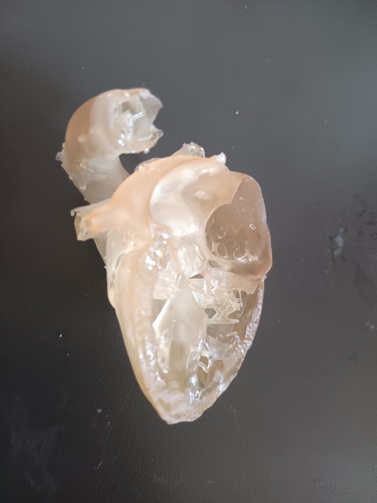
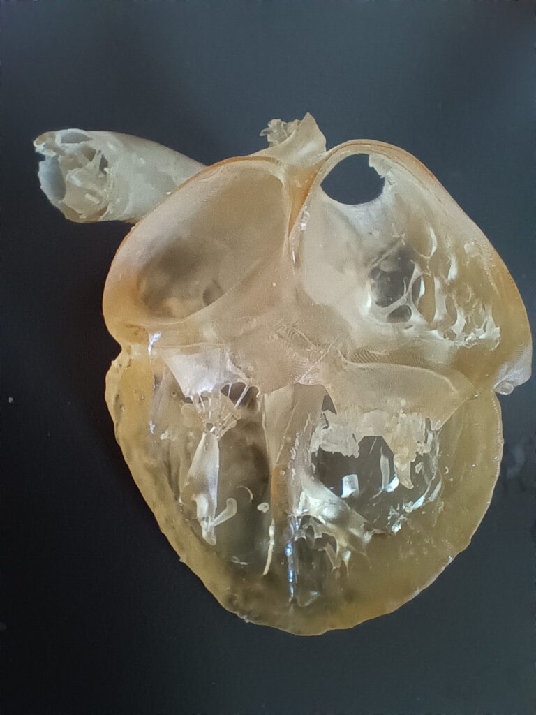
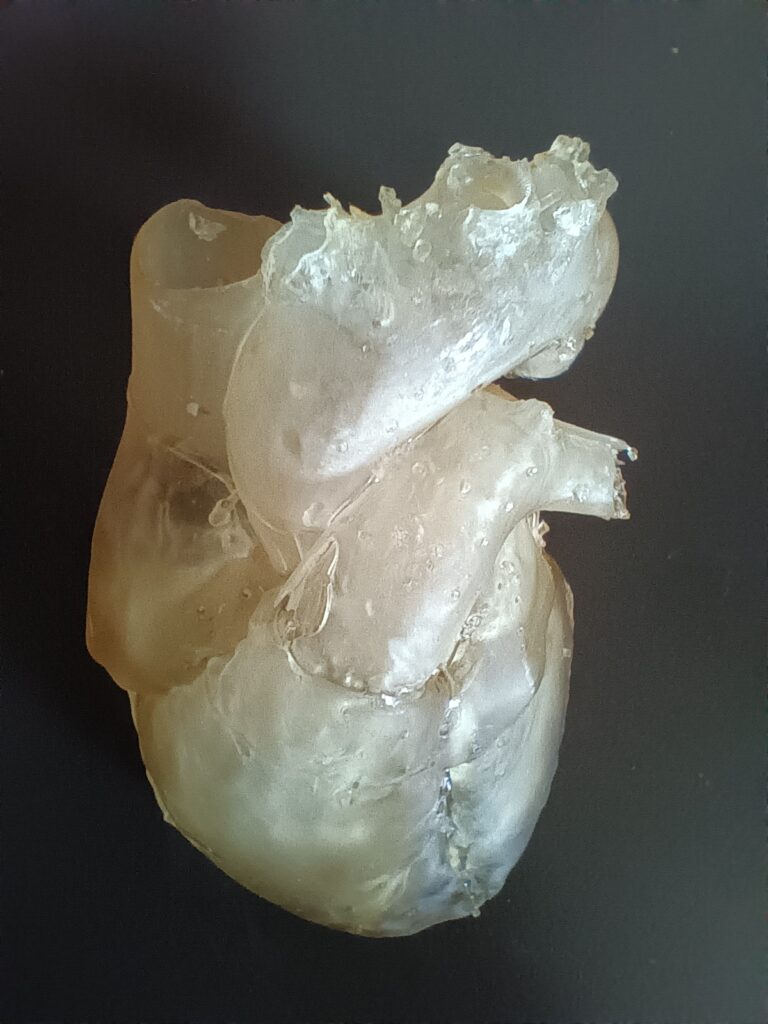
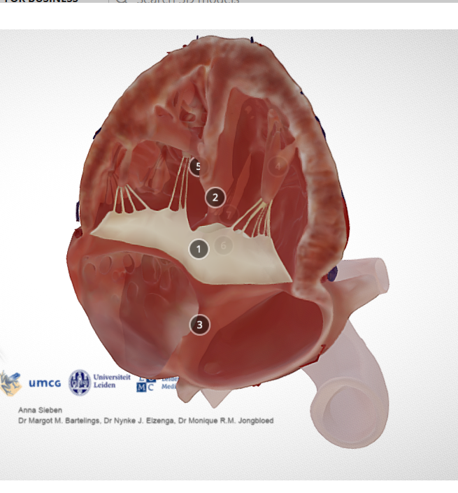
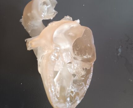
Atrioventricular Septal Defect or (AVSD) can be looked at to explain how the heart functions. Rebecca Howell did this in her STEAM project concerning the defect. Atrioventricular Septal Defect is a congenital heart defect. It is characterized by having a hole in the septa (in between the left and right ventricular wall or atrial wall), while also having faulty valves. The defect is classified under two types incomplete and complete. Atrioventricular Septal Defect is a birth defect that happens in the womb. Babies diagnosed with the defect have an extremely high chance of survival with options such as surgery. The problem with a heart that has an Atrioventricular Septal Defect is that blood can flow between all four chambers, there is only one valve in the center of the heart, and/or the valve is not formed completely causing leakage. Rebecca demonstrated an Atrioventricular Septal Defect by creating a 3D-printed Resin model of the side of a heart. The 3D model has the same characteristics you would see in an Atrioventricular Septal heart including a hole in-between the left and right chambers.