The Growth of Skeletal Muscles after Repeated Exercise of Specific Muscles
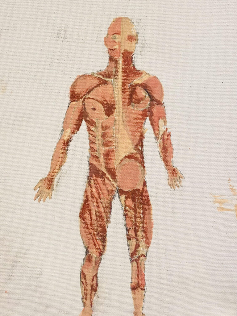
Hello everyone! This is my short summary explaining the image below! I selected the muscles of the skeletal system, and how they change with repeated exercise. This process of change is long and complicated, so we are in for a long ride!
What are Skeletal Muscles? To first understand this process, we must have a solid understanding of the Skeletal Muscle system. The Skeletal Muscles of the body are the largest muscle group in the body, compared to the cardiac and smooth muscle groups, and unlike those groups, exist in most places on the body. Their locations include the length of the appendages, the torso, neck, diaphragm, upper esophagus, and parts of the head including the tongue and eye socket. Skeletal muscles are attached to the bones by tendons, and are composed of muscle fibers, which are surrounded by connective tissue coverings (or sheaths). These connective tissue sheaths include epimysium, perimysium and endomysium. Epimysium tissue is the outermost connective tissue sheath covering an entire muscle organ, and aids in protecting the muscles as well as allowing muscles to contract while maintaining its structure. Perimysium tissue is the connective tissue covering individual muscle fiber bundles, and helps pass on force produced by muscle fibers to the fascicles, which in turn produce muscle contractions. Endomysium tissue sheaths each individual muscle fiber, and allows muscle fibers to slide by each other during contractions with no harm. Skeletal muscle tissue maintains a striated appearance due to the sarcomeres which are different bands of actin and myosin protein myofilaments within the muscle fibers.
How Does Exercise Impact Skeletal Muscle? The skeletal muscles (as previously discussed) are made of sarcomeres which contain two different protein filaments, actin and myosin. Contractions of the skeletal muscle occur when these filaments slide against each other (sliding-filament theory.) The steps in a contraction would be first, the brain begins the process by sending a signal down the spinal cord to the motor neurons which then send their AP (action potential) to the muscle fibers “via neurotransmitter.” The AP then arrives at the axon terminal, causing the “voltage gated calcium channels to open,” making calcium enter the motor neuron. The calcium causes the ACh neurotransmitter to enter the synaptic cleft, making the ACh diffuse “across the ACh receptors,” binding to them. After this, the NMJ axone terminal releases ACh, causing the ACh molecules to diffuse across the synaptic cleft, and bind to the receptors. A “cross bridge” forms between the actin and myosin heads, causing a contraction that lasts as long as Ca++ ions remain in the sarcoplasm to bind to troponin, and as long as ATP is available, “the muscle fiber will continue to shorten.” (Lecture Notes) This process is important to understand in order to comprehend muscle growth over a long period of time.
Muscles can grow in three different ways: “by an increase in cell numbers,” by an increase in “muscle fiber diameter,” and “by an increase in fiber length.” (Am, 1990) However, due to humans being born with almost “full completion” of their multinucleated skeletal muscle cells, after pre and postnatal complete development, there are then only two ways muscles can continue to grow in adulthood. However, before a person is fully developed muscle wise, the muscle cells are enlarging themselves for the increased muscle mass. An increase in “muscle fiber diameter,” occurs when myoblasts are added to the cells, and when myofibrils split into daughter myofibrils, thus causing a greater cross sectional area for muscle fibers. An increase in “muscle fiber length” occurs when new myoblasts are added to the existing muscle fibers, causing an increased amount of nuclei present in the cells. Both of these changes in muscle fibers require “myofibrillar proteins” which are deposited into the cells after being synthesized. It is also suggested that the amount of tension in a muscle controls the amount of “in-series sarcomeres” muscle fibers. (Am 1990) The process of muscle fiber growth induced by weight training is akin to that of muscle hypertrophy, which is a process in which protein synthesis exceeds protein degradation. On a cellular level, this induced muscle growth occurs when the width (or girth) of muscle fibers is increased, rather than the length of the fibers. The increase in myofibrils in muscle fibers can be caused by new stress development which causes an “unequal pressure with splitting at the Z-band and development of additional SR and T-tubule systems.” (Am, 1990) (Phillips, 2009) This means that adding stress on the muscles from training induces myofibril production, causing new development in muscle tissue. Induced skeletal muscle growth can occur when “contractile movements,” and activity, such as strength training, is applied to specific muscles over a period of time. Because muscle mass is only increased when contractions of muscles are pushing or pulling a weighted load, independent growth of muscles implies involuntary, mechanical signaling of some kind. The activation of mTORC1 (mammalian target of rapamycin complex 1), after strength training leads to accelerated protein synthesis, as well as the phosphorylation of two translation regulators: p70S6k and eIF4E, and the binding protein 4E-BP1. Certain hormones also help with muscle development, such as “Androgens, Follistatins, Myostatins, B2 Agonists, Osteocalcin, and IGF-1”, and these hormones and growth factors play a critical role in the physical differences between the muscle size of males and females (with males having significantly greater muscle mass due to a greater natural amount of testosterone, which is a primary androgen.)(Phillips, 2009) Repeated exercise helps not only to shed body fat, but also develop muscles because the repeated stress placed upon skeletal muscles through certain training (strength and resistance training,) if matched with an increase in protein consumption, can lead to increased skeletal muscle mass.
In conclusion, skeletal muscles have the remarkable capability of recreating muscle hypertrophy through an individual’s strength training or hormone consumption. Skeletal muscle, under regulation, can increase in size, and as long as the protein intake exceeds the outtake (metabolism) muscles will recover with added strength and size when met with difficult training.
References:
Am, P. (1990). Muscle growth and exercise. Critical Reviews in Food Science and Nutrition, 29(3), 167–196. https://doi.org/10.1080/10408399009527522
Phillips, S. M. (2009). Physiologic and molecular bases of muscle hypertrophy and atrophy: impact of resistance exercise on human skeletal muscle (protein and exercise dose effects)This paper is one of a selection of papers published in this Special Issue, entitled 14th International Biochemistry of Exercise Conference – Muscles as Molecular and Metabolic Machines, and has undergone the Journal’s usual peer review process. Applied Physiology, Nutrition, and Metabolism, 34(3), 403–410. https://doi.org/10.1139/h09-042