
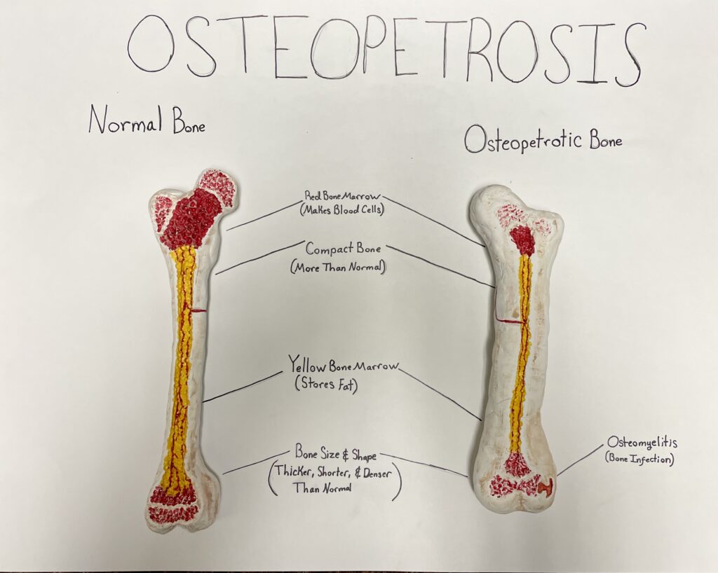
For my STEAM project, I sculpted clay models of bones to show what the skeletal disease osteopetrosis looks like. The course objective I am covering is “know the stages of bone development and repair.” I used air-dry clay to sculpt two femur bones, one that is normal and one with osteopetrosis. The sculptures show both the inside and the outside of each bone. I also included a poster board with my art project that points out some of the key differences between the two bones.
To create my art project, I began by tracing a section of the femur from the Visible Body program. This became the outline for the normal bone sculpture. Next, I used x-ray images of osteopetrotic patients from journal articles to sketch an outline for the osteopetrotic bone. Using these outlines, I sculpted each bone with clay. Once they were dry, I painted on the details, including the medullary cavity containing yellow bone marrow and one of the many blood vessels that supply the bone with blood.
Osteopetrosis is a genetically inherited disorder. There are several different variations of the disease depending on how it is inherited and how severe it is. The disorder causes the osteoclasts in the bone to either not develop fully, or not function properly. The reason the osteoclasts do not work right depends on the variation of osteopetrosis a person has (Stark and Savarirayan, 2009). The osteoclasts are responsible for resorbing, or removing, bone tissue so that the body can use the calcium stored in the bone. When these osteoclasts do not work, osteoblasts can still create more bone tissue, but old bone is not removed. This leads to overly thick and dense bones because the body keeps adding more bone but cannot remove it easily. The large, dense bone tissue of people with osteopetrosis has the appearance of stone, leading this rare disease to be called Marble Bone Disease.
My art piece demonstrates how an osteopetrotic bone differs from a normal bone and shows some of the problems that this disease causes. Osteopetrosis affects all the bones, but I chose to sculpt the femur. I believe this bone most clearly displays osteopetrosis. The most obvious difference between the two clay models is the size and shape. The osteopetrotic bone is thicker, shorter, and less defined than the normal long bone. The amount of compact bone is also much larger for the osteopetrotic bone than the normal bone. Additionally, the distal end of the femur tends to have a more flared shape than a normal femur. The inside of the bones show that the cavity containing bone marrow is very narrow compared to the normal bone. The marrow is where blood cells are produced. Without as much room for bone marrow, not as many blood cells can be created. This causes anemia because of the lack of red blood cells and immune problems because of the lack of white blood cells. One possible outcome of the lack of white blood cells is osteomyelitis. Osteomyelitis is an infection of the bone that causes inflammation and tissue loss in the bones (Stark and Savarirayan, 2009). The pink area inside my bone model represents the osteomyelitis that could happen in a bone that has osteopetrosis.
The issues of osteopetrosis I have listed are common to all bones affected by the disease, but the thickening of some bones creates unique problems. Thickening of the skull shrinks the openings for nerves connecting the eyes and ears to the brain. This puts pressure on these nerves, leading to hearing and seeing problems. Thickening of the skull also reduces blood flow to the brain. This can result in intellectual disability, developmental delay, seizures, and many other neurological conditions. Thickening of the vertebrae can damage the spinal cord (Pillai et al., 2022). Teeth are also affected by this disease. People who have this disease experience tooth eruption later in life because the bone of the jaw is thicker and harder for teeth to move through (Del Fattore et al., 2008).
References
Del Fattore, A., Cappariello, A., & Teti, A. (2008). Genetics, pathogenesis and complications of osteopetrosis. Bone, 42(1), 19–29. https://doi.org/10.1016/j.bone.2007.08.029
Pillai, N. R., Aggarwal, A., & Orchard, P. (2022). Phenotype-autosomal recessive osteopetrosis. Bone, 165. https://doi.org/10.1016/j.bone.2022.116577
Stark, Z., & Savarirayan, R. (2009). Osteopetrosis. Orphanet Journal of Rare Diseases, 4(1). https://doi.org/10.1186/1750-1172-4-5
U.S. Department of Health and Human Services. (2023, August 30). Osteopetrosis. National Institute of Arthritis and Musculoskeletal and Skin Diseases. https://www.niams.nih.gov/health-topics/osteopetrosis
Honor’s project covered osteopetrosis and what it looks like in a bone model, she also compared it to the model of a healthy bone. Osteopetrosis is a bone disorder that causes osteoclasts to either function improperly or not develop fully. The disorder is genetically inherited and it varies in severity and how it’s inherited. When the osteoclasts no longer function properly the body doesn’t reabsorb the bone tissue to use the stored calcium. This then leads to over productive osteoblasts, and bone tissue that is overly dense and looks like stone.