


My project addresses the class objective “Describe the 4 major types of tissues.” This objective is from our Tissues unit, and identifies the four tissue types as epithelial, connective, muscle, and nerve. My project looks at the progression of necrotizing soft tissue infections, such as necrotizing fasciitis and gas gangrene, and how they move through various soft tissues. Necrotizing soft tissue infections all share the characteristics of being caused by bacteria and causing death to the soft tissues of the body such as the epithelial epidermis and dermis, the fascia, the subcutaneous adipose connective tissue, the muscles involved in that part of the body, and even down to the bone. This would also then affect the nervous tissue present in the previously mentioned tissue layers. If it kills the tissue, it also kills the nerves present in that tissue.
Necrotizing soft tissue infections are caused by bacteria that produce toxins leading to tissue destruction, which in turn can lead to sepsis, organ failure, and death, depending on the severity of the infection (1). Necrotizing fasciitis is usually caused by group A Streptococcus or Staphylococcus Aureus. This infection can primarily infect the subcutaneous tissue of the body and its fascia. However, necrotizing fasciitis can also progress so much that it will eventually cause death of the muscle tissue as well (1). Gas Gangrene is another necrotizing soft tissue infection, but is usually caused by Clostridium perfringens. This bacteria is anaerobic and spore forming, leading to the large purple gas bubbles typical to gas gangrene when allowed to progress. This also means that it requires an anaerobic environment in order to develop (4). An infection can begin as necrotizing fasciitis in the outer layers of the skin, fascia, and adipose fat tissue, but it could then progress into gangrene as the blood vessels are destroyed and the blood supply for that whole area of tissues is cut off.
Both necrotizing fasciitis and gas gangrene begin as a great many other tissue infections do, with pain, swelling, fever, edema, and/or erythema (3). They both can stem from a traumatic or non traumatic injury, surgery, or can even be spontaneous (1). Necrotizing fasciitis is often misdiagnosed for other soft tissue infections, such as cellulitis or an abscess (2). But one key feature of necrotizing fasciitis is that the pain felt in the area of infection will be much greater than appears necessary due to the swelling present (3). Necrotizing fasciitis can develop clear blisters/bullae, while gas gangrene will develop large purple bullae. The primary way to diagnose either disease is to perform a variety of blood tests along with bacterial culture tests. Necrotizing fasciitis has what’s called a “finger test.” This is where an incision is made deeply into the infected fascia, and an index finger moved along the tissue plane. If the finger easily dissects the subcutaneous tissue from the fascial plane, then the test is positive. It can also be identified through grey necrotic tissue, destroyed vessels, non contracting muscles, or water, foul smelling fluid called dishwater pus (3). Necrotizing fasciitis and gas gangrene present very similarly along the course of their diseases, except that patients with gas gangrene tend to progress much more rapidly and severely (1).
The treatment for both diseases is very similar; hospitalization is required, as is an extensive course of antibiotics and surgical debridement of the affected tissue. Depending on the bacteria causing the infection, IVIG therapy can be used to reinvigorate the body’s antibodies, and hyperbaric oxygen chamber treatments are used if the bacteria involved is anaerobic (4). Additionally, for both diseases, the severity and progression of infection is greatly affected by any comorbidities the patient might have, such as diabetes, renal failure, liver cirrhosis, or really any other condition seriously affecting the body’s ability to heal itself.
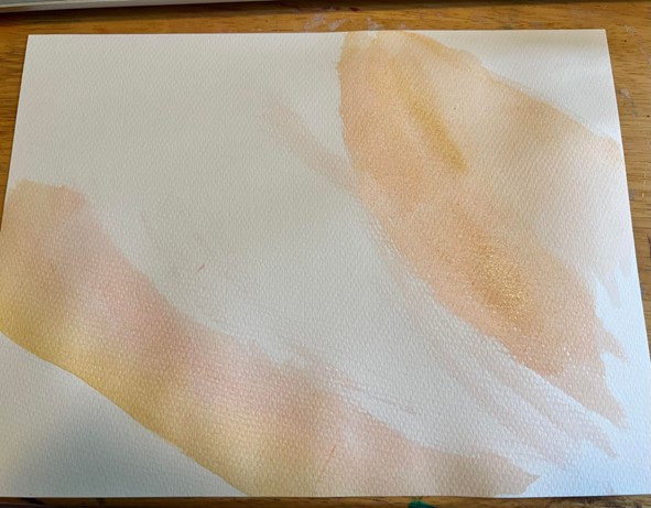
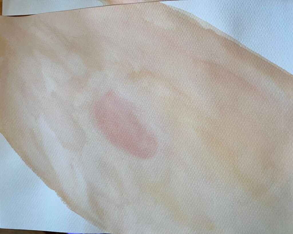
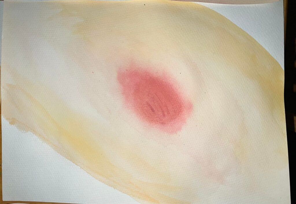
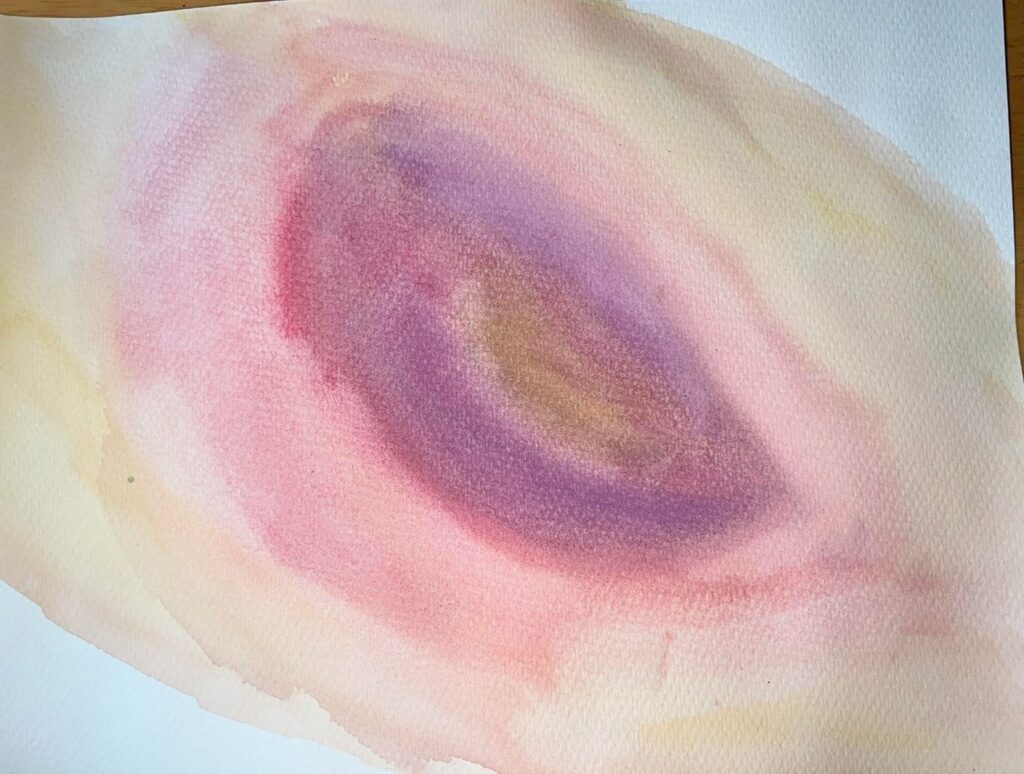
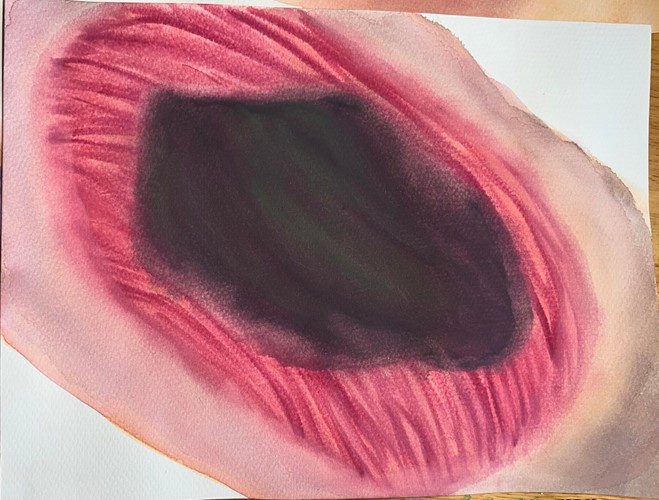
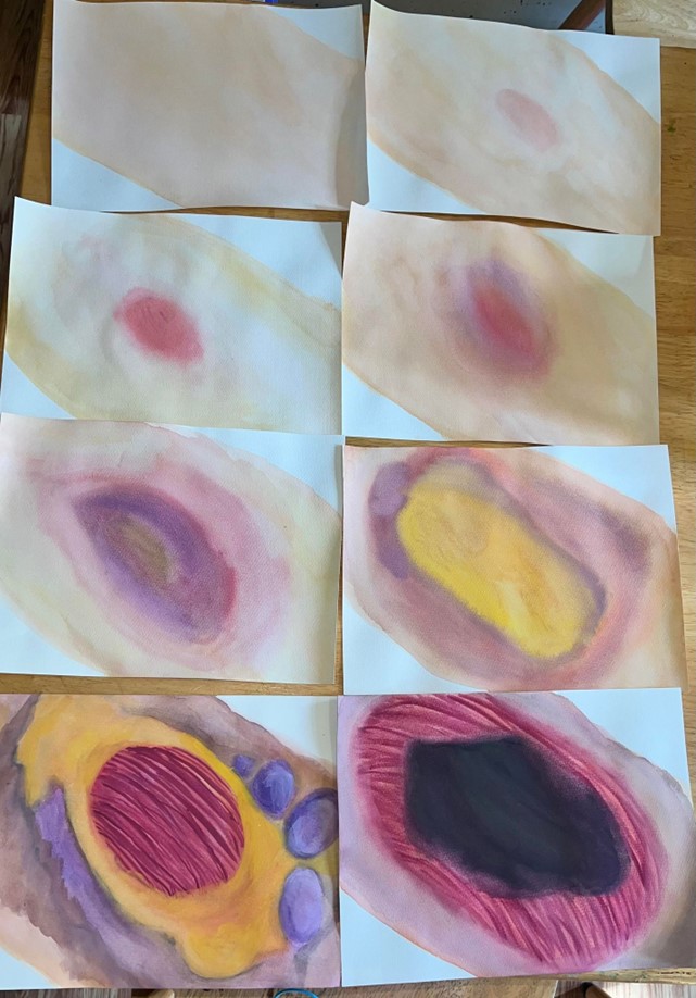
I’ve painted several images displaying the progression of a necrotizing soft tissue infection, starting from regular non infected skin tissue all the way to a severe case. The first image is just a partial painting of the skin to show me painting these. The next three images show a very mild reddening of the skin along with some purple hued swelling and bruising occurring. Image 6 then shows a popped bullae revealing murky dishwater pus underneath. The swelling of the infection has also spread much further around the site. The seventh image shows how the disease has destroyed the initial layers to reveal the yellow adipose tissue plus more severe swelling and spread of infection. The eigth image shows large purple bullae forming along the edges of the infection, greying skin tissue along the edges, additional exposure of adipose tissue, plus the muscle tendons with the fascia destroyed. The final image is meant to portray the tissue after surgical removal of infected tissue and shows where the infections has killed all of the tissue in the center of the image, causing zero blood supply, and exposing the bone. The painted images take some details from necrotizing fasciitis and some from gas gangrene. This is meant to show how many necrotizing soft tissue infections present in a similar way, as well as ending in a similar way. They differ with a few slight characteristics during their progression and in which bacteria needs to be treated.
Sources:
- Leiblein, Maximilian et al. “Clostridial Gas Gangrene – A Rare but Deadly Infection: Case series and Comparison to Other Necrotizing Soft Tissue Infections.” Orthopaedic surgery vol. 12,6 (2020): 1733-1747. doi:10.1111/os.12804
- T Goh and others, Early diagnosis of necrotizing fasciitis, British Journal of Surgery, Volume 101, Issue 1, January 2014, Pages e119–e125, https://doi.org/10.1002/bjs.9371
- Chen, Leon L et al. “Necrotizing fasciitis: A comprehensive review.” Nursing vol. 50,9 (2020): 34-40. doi:10.1097/01.NURSE.0000694752.85118.62
- “Gas Gangrene.” Cleveland Clinic, 14 Feb. 2023, my.clevelandclinic.org/health/diseases/24739-gas-gangrene. Accessed 27 Jul. 2023.
This project looks to assess the effects of necrotizing on the four classes of tissue with the objective of identifying and describing the for major tissue types. Necrotizing soft tissue infections are caused by bacteria that releases toxins that lead to cell death and, if serious enough, organ failure. There are two types of necrotizing infections, necrotizing fasciitis caused by Streptococcus or Staphylococcus Aureus and gas gangrene caused by Clostridium perfringens, but both are very similar. The process of infection as shown starts with a slight reddening of the skin that gets deeper. Purple bruising and swelling begin to appear as the sore worsens. It then shows a popped bullae, bubbles that are caused by the bacterial gas. More of these are shown building up around the sore later. They proceed to show exposed adipose tissue, then exposed muscle tissue. These infections can be caused by any open wound that gets infected or be a spontaneous occurrence. The symptoms such as fever and swelling are similar to other infections, but necrotizing infections are known to be more painful than others. It typically starts at the skin and makes its way down to the muscle and even the bone. If blood vessels are destroyed, then mas cellular death can occur. Testing is done with blood tests, bacterial tests, and a finger test. Once identified, treatment involves hospitalization, surgery, as shown in the final image, and antibiotics. The dead tissue needs to be cut away and what is left needs to be treated.