
The course objective I chose is Know the structure of the knee joint and compare this joint type to other 5 joint types. I wanted to research osteoarthritis and understand what happens to the knee doing this condition. I made two diagrams out of construction paper, one of a healthy knee and another one of a knee with arthritis to show the difference between the two. As shown, the healthy knee has collagen between the femur and the tibia, and the one with arthritis has collagen that has been broken down, causing the two bones to rub against each other creating pain.
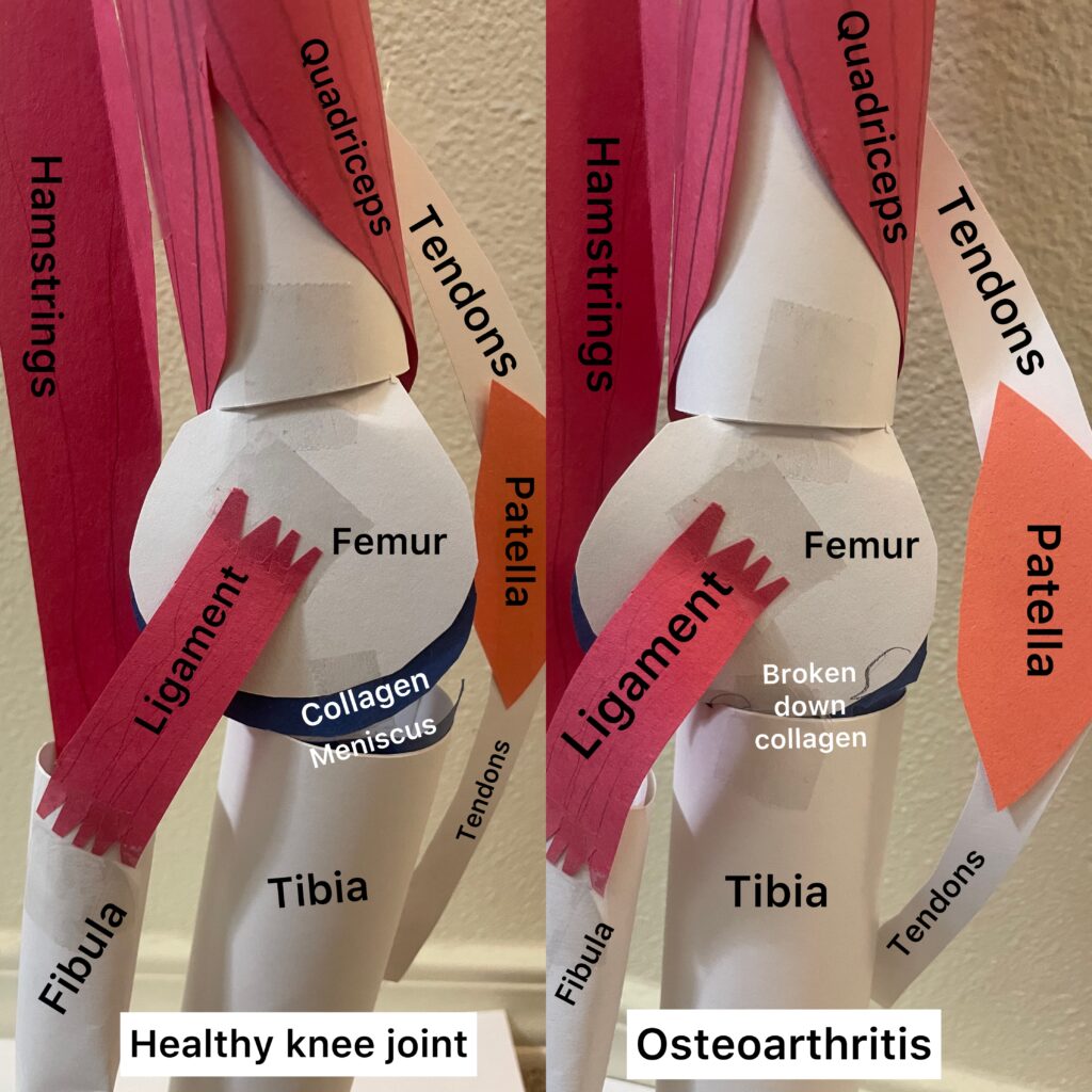
Edward covered the objective of knowing the structure of the knee joint and comparing it to the 5 other types of synovial joints: ball-and-socket, saddle, plane, condylar, and pivot. The knee is classified as a hinge synovial joint, due to its ability for bending and straightening of two bones on a single axis. Ball-and-socket joints are where one bone has a rounded head that fits into a concave articulation on the adjacent bone. Saddle joints are where both bones have a saddle-like shape in different directions. Plane joints are where the bones’ articulating surfaces are flat, slightly curved, of similar size, which allow sliding against each other. Condylar joints are where the bones have a depression at the end that articulates to a rounded structure on an adjacent bone. As for the knee’s structure, the bones that make up the knee are the femur, tibia, and patella. The meniscus is made up of layers of fibrocartilage, there is also hyaline cartilage in the joint. The ligaments involved are the collateral and cruciate ligaments. The patella secured by the quadriceps tendons and patellar ligament. The focus of Edward’s project was how osteoarthritis affects the knee joint. Osteoarthritis causes cartilage in the knee to be damaged and broken down causing friction between the bones, resulting in joint pain and damage to the structures in the knee. The art portion of this project shows all of the structures in the knee and how osteoarthritis causes there to be a breakdown in the meniscus and the collagen between the femur and tibia, causing them to sit on each other.