
Holoprosencephaly
This project is about the complex brain malformation called holoprosencephaly. The objectives covered are “Cleavage, Blastocyst Formation, Implantation, Placental Formation, Gastrulation, Organogenesis, and Birth” and “Describe the major steps of fetal development”. Holoprosencephaly is caused by incomplete cleavage during development. Cleavage is the division of cells during development. Between 18 and 23 days, or during the 5th week, the structure that is developing into a fully formed brain may fail to divide either completely or partially. This can result in severe brain malformation, as well as other midline deformities of the face and skull. These can include cleft palate, single “eye”, lack of nasal development, and others. Holoprosencephaly affects the forebrain, or prosencephalon. These midline facial irregularities are caused because the structures involved are all related to each other during embryonic development. Their precursors are located at the anterior neural crest border and begin to develop together. If there is incomplete separation of the neural tube, these structures are all affected. The nasal cavity and oral cavity undergo significant growth and become two separate whole structures during the fifth week of development; interruption of this can cause these structures to never develop at all, or to partially develop, as seen in the cleft palate, where the oral cavity is left partially open where it had been connected to the nasal cavity. There are several distinct types of holoprosencephaly with varying levels of severity; these are called Alobar, Semilobar, Lobar, Middle Interhemispheric Variant, and Microform. In alobar, there is no or almost no interhemispheric separation. The two halves of the brain remain one, and there is no corpus callosum. As there was no separation and development was entirely interrupted, there are no olfactory bulbs at all and the mouth and nose are replaced with a single cloacal opening along the midline. There may be no eye, or one eye, or some other similar structure. Semilobar is similar to alobar, but there is some separation in the posterior of the brain. This separated area develops the corpus callosum. There are no or severely underdeveloped olfactory bulbs, and the nasal and oral structures are still fused though they may be more developed. In lobar holoprosencephaly, most of the brain is separated into hemispheres, between which develops most of a corpus callosum. MIHV is similar to lobar, but the nonseparation is in the posterior frontal and parietal lobes. In microform, the brain has entirely separated into its two hemispheres, but there may still be some subtle defects along the midline of the brain or face. These can include intellectual disability, movement disorders, autonomic dysfunction, and epilepsy. Fetuses with alobar, semilobar, and MIHV holoprosencephaly are often nonviable. Those that do tend to have shorter lives. Even those with less severe forms often require lifelong treatment and medication. There are several risk factors for this condition, such as drug use or diabetes of the mother during pregnancy, as well as some genetic factors. But there is no guaranteed way to predict or prevent it.
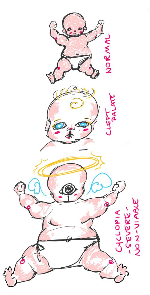
My art project shows 3 babies, one without holoprosencephaly, one with mild microform holoprosencephaly (cleft palate), and one with a severe form of holoprosencephaly showing a single cloacal facial slit and singular eye structure- I included this structure as it is the origin of holoprosencephaly’s more common name, cyclopia. The angel symbology represents its lack of viability, though I have drawn it as a plump full term baby to make the structural differences more obvious. This digital image was drawn on a Microsoft surface in OneNote with a surface pen.
References
Raam, M. S., Solomon, B. D., & Muenke, M. (2011). Holoprosencephaly: a guide to diagnosis and clinical management. Indian pediatrics, 48(6), 457–466. https://doi.org/10.1007/s13312-011-0078-x
Som, P. M., & Naidich, T. P. (2013). Illustrated Review of the embryology and development of the facial region, part 1: Early face and lateral nasal cavities. American Journal of Neuroradiology, 34(12), 2233–2240. https://doi.org/10.3174/ajnr.a3415
Dubourg, C., Bendavid, C., Pasquier, L. et al. Holoprosencephaly. Orphanet J Rare Dis 2, 8 (2007). https://doi.org/10.1186/1750-1172-2-8
I really like your artwork, the angel imagery is fitting and creative. The color pallet is pleasing to the eye. The close up shot of the baby with the cleft palate is really well done and the placement of the skin tones is very effective. The overall sketchy style works well for the idea, it adds to the ethereal nature of the third baby.
I’ve seen some imagery of this, but I didn’t know it was caused by holoprosencephaly. It is very interesting to look at as you can see where the facial features develop and how the parts migrate during development. The most notable to me is the presence of the fleshy proboscis above the eye sockets when they aren’t divided. On the severe side of the spectrum, most fetuses are non viable and a large number that are born die within the first six months. While those with very mild holoprosencephaly can be non-symptomatic and live unaffected by the condition.