

This STEAM Project is about Temporomandibular Disorders, or TMDs. These disorders are often called TMJ, however, this term properly only refers to the associated Temporomandibular joint. The temporomandibular joint is a synovial joint containing an articular disc. This joint connects the mandible and the temporal bone. Movement of this joint allows actions such as talking and chewing. TMDs are a group of disorders with three main classes (National Institute of Dental and Craniofacial Research, 2022), which are disorders of the temporomandibular joint, the muscles of mastication, or TMD specific headaches. The unit objectives connected to this project are ”Know the list of bones provided in blackboard” and “Know the structure of the knee joint and compare this joint type to other 5 joint types”. Assessment for TMDs is done using the Diagnostic Criteria for TMD (DC/TMD), which allows for multiple diagnoses (Ohrbach, 2016). For the purposes of this project, I have contained the scope of it to just joint disorders and diseases.
The most common TMD is Anterior Disc Displacement (Dalewski, 2020). In this condition, the disc becomes displaced towards the mandibular notch and can become deformed or torn. Another common TMD is osteoarthritis of the temporomandibular joint. This can be caused by acute injury, long-term stress on the joint, or associated disease. In osteoarthritis, loss or damage to the cartilage in the joint causes stress and wear on the bone and this in turn can cause changes in the shape of the overall structure and function of the joint (Kalladka, 2013). Some common changes to the shape of the mandibular condyle include “cane-shaped remodeling”, pole depression or flattening, surface erosion, and bone spurs (Nah, 2012).
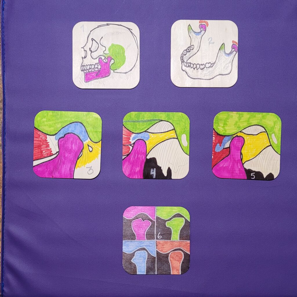
My project consists of six wooden tiles with illustrations of some anatomical structures and issues associated with TMD. Materials used are wooden tiles, pencil, permanent marker, and paint pen.
Tile 1 is the skull, with the temporal bone, mandible, and temporomandibular joint highlighted.
Tile 2 is the mandible, with the condylar process, the coronoid process, mandibular notch, and temporomandibular joint highlighted.
Tile 3 is a healthy mandibular joint with the temporal bone (specifically the zygomatic arch), the condylar process of the mandible, the lateral pterygoid muscle, articular disc, and ligaments highlighted.
Tile 4 shows the same structures highlighted as tile 3, but with the most common TMD, Anterior Disc Displacement. There are two types of this TMD, with Reduction and without. This tile shows with Reduction. In this TMD, the disc slides out of place when the jaw is opened (Dalewski, 2020).
Tile 5 shows Anterior Disc Displacement without Reduction. In this TMD, the disc slides further out of place and becomes stuck in the anterior joint recess, causing severe dysfunction of the temporomandibular joint (Dalewski, 2020). Tiles 3, 4 and 5 were based on images and descriptions from the Dalewski article.
Tile 6 shows four common physical changes to the mandibular condyles that occur with osteoarthritis, a common TMD. These illustrations are based on cone beam computed tomography (CBCT) imaging from the 2012 Nah article.
U.S. Department of Health and Human Services. (2022, January). TMD (temporomandibular disorders). National Institute of Dental and Craniofacial Research. Retrieved November 27, 2022, from https://www.nidcr.nih.gov/health-info/tmd
Ohrbach, R., & Dworkin, S. F. (2016). The Evolution of TMD Diagnosis: Past, Present, Future. Journal of dental research, 95(10), 1093–1101. https://doi.org/10.1177/0022034516653922
Dalewski, B., Kamińska, A., Białkowska, K., Jakubowska, A., & Sobolewska, E. (2020). Association of Estrogen receptor 1 and tumor necrosis factor α polymorphisms with temporomandibular joint anterior disc displacement without reduction. Disease Markers, 2020, 1–9. https://doi.org/10.1155/2020/6351817
Kalladka, M., Quek, S., Heir, G., Eliav, E., Mupparapu, M., & Viswanath, A. (2013). Temporomandibular joint osteoarthritis: Diagnosis and long-term conservative management: A topic review. The Journal of Indian Prosthodontic Society, 14(1), 6–15. https://doi.org/10.1007/s13191-013-0321-3
Nah, K.-S. (2012). Condylar bony changes in patients with temporomandibular disorders: A CBCT study. Imaging Science in Dentistry, 42(4), 249. https://doi.org/10.5624/isd.2012.42.4.249
This STEAM Project is about Temporomandibular Disorders, or TMDs. These disorders are often called TMJ, however, this term properly only refers to the associated Temporomandibular joint. The temporomandibular joint is a synovial joint containing an articular disc. This joint connects the mandible and the temporal bone. Movement of this joint allows actions such as talking and chewing. TMDs are a group of disorders with three main classes (National Institute of Dental and Craniofacial Research, 2022), which are disorders of the temporomandibular joint, the muscles of mastication, or TMD specific headaches. The unit objectives connected to this project are ”Know the list of bones provided in blackboard” and “Know the structure of the knee joint and compare this joint type to other 5 joint types”. Assessment for TMDs is done using the Diagnostic Criteria for TMD (DC/TMD), which allows for multiple diagnoses (Ohrbach, 2016). For the purposes of this project, I have contained the scope of it to just joint disorders and diseases.
The most common TMD is Anterior Disc Displacement (Dalewski, 2020). In this condition, the disc becomes displaced towards the mandibular notch and can become deformed or torn. Another common TMD is osteoarthritis of the temporomandibular joint. This can be caused by acute injury, long-term stress on the joint, or associated disease. In osteoarthritis, loss or damage to the cartilage in the joint causes stress and wear on the bone and this in turn can cause changes in the shape of the overall structure and function of the joint (Kalladka, 2013). Some common changes to the shape of the mandibular condyle include “cane-shaped remodeling”, pole depression or flattening, surface erosion, and bone spurs (Nah, 2012).
My project consists of six wooden tiles with illustrations of some anatomical structures and issues associated with TMD. Materials used are wooden tiles, pencil, permanent marker, and paint pen.
Tile 1 is the skull, with the temporal bone, mandible, and temporomandibular joint highlighted.
Tile 2 is the mandible, with the condylar process, the coronoid process, mandibular notch, and temporomandibular joint highlighted.
Tile 3 is a healthy mandibular joint with the temporal bone (specifically the zygomatic arch), the condylar process of the mandible, the lateral pterygoid muscle, articular disc, and ligaments highlighted.
Tile 4 shows the same structures highlighted as tile 3, but with the most common TMD, Anterior Disc Displacement. There are two types of this TMD, with Reduction and without. This tile shows with Reduction. In this TMD, the disc slides out of place when the jaw is opened (Dalewski, 2020).
Tile 5 shows Anterior Disc Displacement without Reduction. In this TMD, the disc slides further out of place and becomes stuck in the anterior joint recess, causing severe dysfunction of the temporomandibular joint (Dalewski, 2020). Tiles 3, 4 and 5 were based on images and descriptions from the Dalewski article.
Tile 6 shows four common physical changes to the mandibular condyles that occur with osteoarthritis, a common TMD. These illustrations are based on cone beam computed tomography (CBCT) imaging from the 2012 Nah article.
**Please disregard the previous comment- posted in error**
Temporomandibular Disorders, or TMDs are disorders are often called TMJ, however, this term properly only refers to the associated Temporomandibular joint. The temporomandibular joint is a synovial joint containing an articular disc. This joint connects the mandible and the temporal bone. Movement of this joint allows talking and chewing. TMDs are a group of disorders which are disorders of the temporomandibular joint, the muscles of mastication, or TMD specific headaches.
The most common TMD is Anterior Disc Displacement. In this condition, the disc becomes displaced towards the mandibular notch and can become deformed or torn. Another common TMD is osteoarthritis of the temporomandibular joint. This can be caused by acute injury, long-term stress on the joint, or associated disease.