
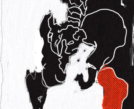
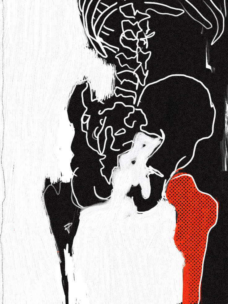
This project’s purpose is to explore the process of diagnosing osteoporosis. I will first introduce osteoporosis and its effects, then I will cover the bone cells and their roles in bone building and resorption to understand how osteoporosis works, and finally the imaging process. My art piece on osteoporosis aims to communicate the fragile and deteriorative nature of the disease. The black background and the skeleton’s outline are meant to resemble an x-ray scan. The line work is kept natural to highlight instability. The red bone represents an osteoporotic bone.
Osteoporosis is a disease characterized by deteriorating bone volume and low density resulting in a stronger likelihood of suffering a fragility fracture with increasing relevance amongst the population. An estimated 200 million may have osteoporosis with numbers likely to increase as the population becomes older. Developing osteoporosis in a lifetime is exponential with age although it can affect any age group. Osteoporosis is not usually detected until a fragility fracture has occurred; it is primarily asymptomatic until the osteoporotic bone encounters low-force trauma. It is also a disease that is disruptive in other aspects of life. Suffering from a fragility fracture on the hip can cause disability, rehabilitation, and financial burden.
The cause of Osteoporosis has many factors, they are grouped into type I and type II. Type I is caused by a lack of estrogen. Loss of estrogen is associated with higher bone resorption rates because estrogen reduces the amount of osteoclast differentiation, cells responsible for bone breakdown (Väänänen, 1996). Type II is osteoporosis caused by inadequate amounts of calcium; causes of this are inhibited absorption of calcium either from an illness preventing the body from absorbing calcium or from inefficient amounts of vitamin D that aid in calcium absorption. Activation of parathyroid hormone (PTH) also falls into type II osteoporosis. PTH is responsible for activating osteoclasts when the body detects low amounts of calcium in the blood. When osteoclasts activate, they break down calcium from bones and return them to the bloodstream. Achieving sufficient peak bone mass is a crucial factor in preventing severe bone loss later in life. Peak bone mass is attained at around the age of 30, then a loss of bone mass at a rate of 0.3% per year or 2% in menopausal women sets in; even without compromise to bone loss rates, having an insufficient peak bone mass still means a likelihood of developing osteoporosis (Dobbs, 1999). Factors preventing the build of peak bone mass at an early age include immobilization for extended periods of time, eating disorders, illness, or genetic predisposition.
Patients with fractures and are over the age of 50, women over 60, or patients with a body weight less than 154.3 lb./ 70 kg should be screened for osteoporosis. A blood test may be done to check for secondary causes of osteoporosis. Bone densitometry tests gauge patients based on bone mineral density on whether they have normal or osteoporotic bone, it also predicts the risk of bone fractures. Body mineral density (BMD) is used as a close indicator of bone strength, it is obtained using a dual-energy x-ray absorptiometry scan (DXA). A DXA scan uses harmless doses of radiation to create an image of bones with no invasive procedures necessary. Technicians will take images of the lumbar or thoracic spine and femoral neck and use them to determine BMD. BMD is then used to determine T-score and Z-score. A T-score is a gap between an individual BMD and the average BMD of a 20 to 40-year-old. Having a T-score lower than –2.5 is an indication of osteoporosis. -2.5 to –1.0 indicates osteopenia or lower than average BMD. Z-score is the gap between an individual BMD and the average BMD of the same age group. 0 is normal BMD and Z-scores below –2.0 is considered osteoporotic. The risk of a fracture is assessed using FRAX, an algorithm that uses an individual’s age, sex, and body mass index (Kanis, 2011).
References
Kanis, J.A., Hans, D., Cooper, C. et al. Interpretation and use of FRAX in clinical practice. Osteoporos Int 22, 2395–2411 (2011). https://doi.org/10.1007/s00198-011-1713-z.
López-López J, Estrugo-Devesa A, Jane-Salas E, Ayuso-Montero R, Gómez-Vaquero C. Early diagnosis of osteoporosis by means of orthopan . Early diagnosis of osteoporosis by means of orthopantomograms and oral x-rays: A systematic review. Med Oral Patol Oral Cir Bucal. 2011 Nov 1;16 (7):e905-13. http://www.medicinaoral.com/medoralfree01/v16i7/medoralv16i7p905.pdf.
RadiologyInfo. 2022. In Bone Density Scan (DXA or DEXA).
Pisani P., Renna M.D., Conversano F., Casciaro E., Muratore M., Quarta E., Paola M.D., Casciaro S. Screening and early diagnosis of osteoporosis through X-ray and ultrasound based techniques. World J Radiol. 2013 Nov 28;5(11):398-410. doi: 10.4329/wjr.v5.i11.398. PMID: 24349644; PMCID: PMC3856332.
Nelson D., Peterson E., Tilley B., Otallon W.M., Chao E., Riggs B.L., Kleerekoper M. Measurement of vertebral area on spine X-rays in osteoporosis: Reliability of digitizing techniques. 2022 American Society for Bone and Mineral Research. https://doi.org/10.1002/jbmr.5650050707.
Rachner T.D., Khosla S., Hofbauer L.C. Elsevier (2011) .Osteoporosis: now and the future.
Robert M., David F., David W.D., Marjorie L., Jane A.C. (June 13, 2013). Osteoporosis.
Sözen T, Özışık L, Başaran NÇ. An overview and management of osteoporosis. Eur J Rheumatol. 2017 Mar;4(1):46-56. doi: 10.5152/eurjrheum.2016.048. Epub 2016 Dec 30. PMID: 28293453; PMCID: PMC5335887.
Riggs BL. Overview of osteoporosis. West J Med. 1991 Jan;154(1):63-77. PMID: 2024511; PMCID: PMC1002677.
Dobbs MB, Buckwalter J, Saltzman C. Osteoporosis: the increasing role of the orthopaedist. Iowa Orthop J. 1999;19:43-52. PMID: 10847516; PMCID: PMC1888612.
Heaney, R. P., Abrams, S., Dawson-hughes, B., Looker, A., Marcus, R., Matkovic, V., & Weaver, C. (2000). Peak bone mass. Osteoporosis International, 11(12), 985-1009. doi:https://doi.org/10.1007/s001980070020.
Väänänen HK, Härkönen PL. Estrogen and bone metabolism. Maturitas. 1996 May;23 Suppl:S65-9. doi: 10.1016/0378-5122(96)01015-8. PMID: 8865143.
Riggs B.L., Jowsey J., Kelly P.J, Jones J.D., Maher F.T. Affects of Hormones on Bone in Primary Osteoporosis. J Clin Invest. 1969;48(6):1065-1072. https://doi.org/10.1172/JCI106062.
Nazrun AS, Tzar MN, Mokhtar SA, Mohamed IN. A systematic review of the outcomes of osteoporotic fracture patients after hospital discharge: morbidity, subsequent fractures, and mortality. Ther Clin Risk Manag. 2014 Nov 18;10:937-48. doi: 10.2147/TCRM.S72456. PMID: 25429224; PMCID: PMC4242696.
Bone Densitometry. https://www.hopkinsmedicine.org/health/treatment-tests-and-therapies/bone-densitometry.
Lorentzon M., Cummings S.R. Osteoporosis: The Evolution of a Diagnosis. Journal of Internal Medicine. https://doi.org/10.1111/joim.12369.
Cummings SR, Bates D, Black DM. Clinical Use of Bone Densitometry: Scientific Review. JAMA. 2002;288(15):1889–1897. doi:10.1001/jama.288.15.1889.
U.S Preventative Task Force. (2002). Screening for Osteoporosis in Postmenopausal Women. Agency for Healthcare Research and Quality Pub. No. 03-511A.
Adler, R. Osteoporosis in men: a review. Bone Res 2, 14001 (2014). https://doi.org/10.1038/boneres.2014.1.
Jox Iways STEAM Project was Diagnosing Osteoporosis, the objective covered was Identifying bone cells and the roles they had. Osteoporosis is the loss of bone volume and low density this causes the bone to become more fragile and more likely to break. The probability of diagnosing osteoporosis is low without a break in the bone first as it goes on to be asymptomatic until the bone encounters low force trauma. Osteoporosis has two types. Type I is caused by the lack of estrogen. Lack of Estrogen causes osteoclast to increase because estrogen helps keep all the bone cells in balance and when there is a lack of estrogen then osteoclast will start absorbing the bone with not enough osteoblast to build the bone back. Type II is caused by deficiency of calcium. Diagnosing of osteoporosis starts when you have a fracture. Patients who are over 50, women over 60, or people wIth low body weight should be screened if they have a bone fracture. Bone densitometry test if the mineral density of the bone is normal or not and figures out how likely you are to fracture your bone. The way of screening for BMD is a dual energy x-ray Absorptiometry scan. This is then scored using a T-score and a Z-score. The T-score is the gap between a patients BMD and the average BMD of a 20–40-year-old, lower the -2.5 is an indication of osteoporosis. Z-Score is the gap between a patient BMD and the average BMD of the same age group, anything below -2.0 is considered osteoporotic.