The course objective I chose is to explain the structure and function of the heart, as well as explain a Ventricular Septal Defect. The heart is a discrete, localized pumping structure around the size of a fist. It pumps blood through the entire body and is made up of many layers of tissue. It contains blood vessels including veins, capillaries, and arteries, each of which transports blood to the body. It holds the atria or the receiving chambers, which are small, thin-walled chambers that contribute very little to the propulsion of blood. The ventricles, or the discharging chambers, are the actual pumps of the heart and they make up most of the volume of the heart. The atrioventricular (AV) valves prevent backflow into the atria when the ventricles contract. When the AV valves are open, the atrial pressure is greater than the ventricular pressure. When the AV valves are closed, the atrial pressure is less than the ventricular pressure. The semilunar SL valves prevent backflow from major arteries back into the ventricles.
The heart defect I’ll be covering is the ventricular septal defect or VSD. It’s one of the most common congenital heart defects and makes up for roughly 40% of all heart abnormalities. (Penny & Vick lll, 2011). In a healthy heart, the right ventricle pumps blood into the pulmonary trunk and the left ventricle pumps blood into the aorta. However, in a VSD, the oxygenated blood flows from the left ventricle to the right ventricle due to a hole in the heart located in the ventricular septum. (Mayo Clinic Staff, 2021). The ventricular septum is the wall that separates the heart’s lower chambers. In turn, when the septum has a hole in it, it allows blood to pass from the left to the right side of the heart. So when the blood is flowing to the right ventricle rather than the aorta, the oxygenated blood is then pumped back to the lungs instead of to the body, forcing the heart to work much harder than normal. (Minette & Sahn, 2006).
There are quite a few factors that could lead to the development of a ventricular septal defect. Possibilities that could include the interventricular septum failing to form completely during fetal development. It’s also possible that the atrioventricular cushions failed to fuse with the septum or each other, and lastly, the area that makes the membraneous septum fails to close entirely. VSD as well as most other congenital heart abnormalities often have multifactorial origins and many people with Ventricular Septal Defects are asymptomatic. However, some people can experience difficulty breathing abnormal skin color, and just overall abnormal breathing. There are few treatment options, however, most patients require subacute bacterial endocarditis prophylaxis, while there are other catheter, surgery, or a combination of both options. (Penny & Vick lll, 2011).
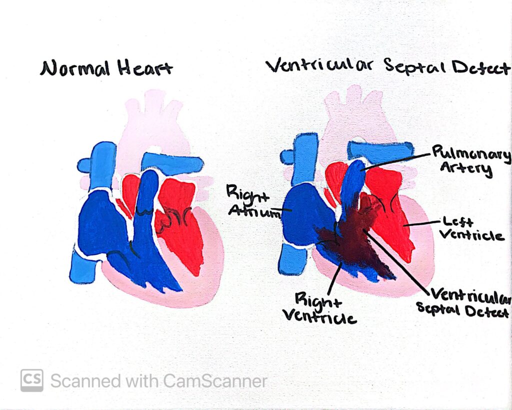
In the painting, I’ve displayed a picture of a healthy heart with a normal ventricular septum. Alongside the healthy heart, is a heart containing a ventricular septal defect with the hole in the septum allowing blood to pass from the left ventricle into the right.
Emily Stacy
This project is shown to explain the function and structure of the heart. The disorder/defect they decided to talk about is the Ventricular Septal Disorderor VSD. The heart’s function is to pump blood through the whole body and is made of many different layers of tissue. The ventricles are actually the heart’s pumps and make up most of the heart’s volume. The atrioventricular valves prevent backflow into the atria when the ventricles contract when pumping. Then the semilunar valves prevent the backflow of blood from major arteries into the ventricles.
VSD is a disorder of the heart where oxygenated blood flows from the left ventricle to the right due to a hole in the septum. This disorder is the most common type of congenital heart defect and people can experience difficulty breathing, abnormal breathing, and abnormal skin colors. This disorder makes up roughly 40% of all heart abnormalities as well. The paintings linked in this STEAM project show two different hearts, normal health, a working heart, and a heart affected by VSD.

The course objective I chose is to explain the structure and function of the heart, as well as explain a Ventricular Septal Defect. The heart is a discrete, localized pumping structure around the size of a fist. It pumps blood through the entire body and is made up of many layers of tissue. It contains blood vessels including veins, capillaries, and arteries, each of which transports blood to the body. It holds the atria or the receiving chambers, which are small, thin-walled chambers that contribute very little to the propulsion of blood. The ventricles, or the discharging chambers, are the actual pumps of the heart and they make up most of the volume of the heart. The atrioventricular (AV) valves prevent backflow into the atria when the ventricles contract. When the AV valves are open, the atrial pressure is greater than the ventricular pressure. When the AV valves are closed, the atrial pressure is less than the ventricular pressure. The semilunar SL valves prevent backflow from major arteries back into the ventricles.
The heart defect I’ll be covering is the ventricular septal defect or VSD. It’s one of the most common congenital heart defects and makes up for roughly 40% of all heart abnormalities. (Penny & Vick lll, 2011). In a healthy heart, the right ventricle pumps blood into the pulmonary trunk and the left ventricle pumps blood into the aorta. However, in a VSD, the oxygenated blood flows from the left ventricle to the right ventricle due to a hole in the heart located in the ventricular septum. (Mayo Clinic Staff, 2021). The ventricular septum is the wall that separates the heart’s lower chambers. In turn, when the septum has a hole in it, it allows blood to pass from the left to the right side of the heart. So when the blood is flowing to the right ventricle rather than the aorta, the oxygenated blood is then pumped back to the lungs instead of to the body, forcing the heart to work much harder than normal. (Minette & Sahn, 2006).
There are quite a few factors that could lead to the development of a ventricular septal defect. Possibilities that could include the interventricular septum failing to form completely during fetal development. It’s also possible that the atrioventricular cushions failed to fuse with the septum or each other, and lastly, the area that makes the membraneous septum fails to close entirely. VSD as well as most other congenital heart abnormalities often have multifactorial origins and many people with Ventricular Septal Defects are asymptomatic. However, some people can experience difficulty breathing abnormal skin color, and just overall abnormal breathing. There are few treatment options, however, most patients require subacute bacterial endocarditis prophylaxis, while there are other catheter, surgery, or a combination of both options. (Penny & Vick lll, 2011).
In the painting, I’ve displayed a picture of a healthy heart with a normal ventricular septum. Alongside the healthy heart, is a heart containing a ventricular septal defect with the hole in the septum allowing blood to pass from the left ventricle into the right.
This project is shown to explain the function and structure of the heart. The disorder/defect they decided to talk about is the Ventricular Septal Disorderor VSD. The heart’s function is to pump blood through the whole body and is made of many different layers of tissue. The ventricles are actually the heart’s pumps and make up most of the heart’s volume. The atrioventricular valves prevent backflow into the atria when the ventricles contract when pumping. Then the semilunar valves prevent the backflow of blood from major arteries into the ventricles.
VSD is a disorder of the heart where oxygenated blood flows from the left ventricle to the right due to a hole in the septum. This disorder is the most common type of congenital heart defect and people can experience difficulty breathing, abnormal breathing, and abnormal skin colors. This disorder makes up roughly 40% of all heart abnormalities as well. The paintings linked in this STEAM project show two different hearts, normal health, a working heart, and a heart affected by VSD.