
For my STEAM project, I will be talking about a rare congenital heart condition known as Tetralogy of Fallot (“fuh-loh”). The objectives I’ll be covering are “know the path of blood through the heart and circulatory system” and “explain the structure and function of the heart”. I chose this topic because when I was born, I was diagnosed with the condition. The doctors kept me in the hospital for two months to monitor my heart and fortunately for me, my heart reduced to normal size and function.
Tetralogy of Fallot (TOF) is a combination of four defects that disrupt the flow of the heart, thus the name contains “tetra” meaning four. Etienne-Louis Arthur Fallot coined the term “tetralogy” in 1888, in whom TOF got its name. Contrary to being a rare condition, TOF is the most common cause of cyanotic cardiac disease in patients beyond neonatal age. The cause of cyanosis is due to the malfunction of the structures in the heart.
In normal circulation, unoxygenated blood from the body is transported to the right side of the heart—right atrium to right ventricle—via the vena cava where it is then pumped to the pulmonary artery and to the lungs to become oxygenated. From there, the pulmonary veins transport the oxygenated blood from the lungs to the left side of the heart—left atrium to left ventricle—to be pumped into the aorta and distributed throughout the body. Patients born with TOF do not have normal circulation because they are presented with four major structural defects: ventricular septal defect, an overriding aorta, pulmonary stenosis, and hypertrophy of the right ventricle.
A ventricular septal defect (VSD) is a birth defect in which there is a hole in the septum (wall) between the two lower chambers of the heart; the ventricles. It occurs when the septum does not fully develop. As a result, VSD allows oxygenated blood to mix with unoxygenated blood and flow through both the pulmonary artery and the aorta.
Normally, the aorta attaches to the left ventricle which pumps oxygenated blood through it. In an overriding aorta, the aorta is displaced directly over the VSD which allows the unoxygenated blood from the right ventricle to pour into the left ventricle and flow straight into the aorta where it enters systemic circulation.
Pulmonary stenosis (narrowing) occurs when the pulmonary artery—the blood vessel that delivers unoxygenated blood from the right ventricle to the lung—is narrowed which prevents the pulmonary valve from opening completely. This results in less blood reaching the lungs, causing an accumulation of unoxygenated blood. The degree of stenosis can be exacerbated by catecholamines, or a state of low intravascular volume, which increases the patients susceptibility to sudden and acute episodes of desaturation known as hypercyanotic spells. Since blood takes the path of least resistance and the pulmonary artery is restricted due to stenosis, the unoxygenated blood flows easily through the VSD into the overriding aorta and through systemic circulation. The baby then becomes cyanotic because there is insufficient oxygenated blood present.
Right ventricular hypertrophy is the thickening of the right ventricle muscular wall. This occurs because the right ventricle must work harder to pump blood through the stenosed (narrowed) pulmonary valve. This thickened wall gives the heart a boot-like appearance which is one of TOF’s more easily recognizable characteristics for diagnoses.
The cause of TOF is unknown but it can be treated through surgery or a palliative (temporary) shunt. Most babies and children have open-heart surgery known as intracardiac repair to correct the defects. In an intracardiac repair procedure, the VSD is patched over to seal the hole between the ventricles. The stenosed pulmonary valve is repaired or replaced to increase blood circulation to the lungs. In some cases, babies must undergo a palliative surgery prior to intracardiac repair; also to improve blood flow to the lungs. This procedure is typically done if the baby was born prematurely or has underdeveloped pulmonary arteries (hypoplastic). In the procedure, a shunt (bypass) is created between a large artery that branches off from the aorta and the pulmonary artery. When the baby is ready for intracardiac repair, the shunt is removed during the procedure.
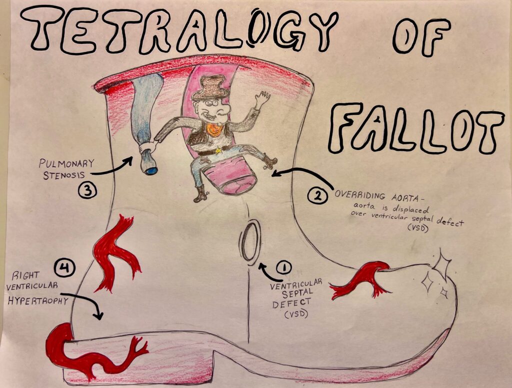
Lynciemae’s piece describes tetralogy of fallot. Tetralogy of fallot is a rare condition caused by a combination of four rare defects that are present at birth. Tetralogy of Fallot defects cause oxygen-poor blood to flow out of the heart and into the rest of the body. In patients with normal circulation, unoxygenated blood from the body is transported to the right side of the heart. It then flows from the right atrium to the right ventricle. Finally, it flows to the vena cava where it will be pumped to the pulmonary artery to go onto the lungs to be oxygenated. After being oxygenated, the pulmonary veins will transport the oxygenated blood from the lungs to the left side of the heart, to the left atrium followed by the left ventricle. It will then be pumped to the aorta to be given to the rest of the body. Ventricular septal defect (VSD) is a birth defect where there is a hole in the septum between the two lower chambers of the heart which develops when the septum does not fully close, allowing oxygenated blood to mix with deoxygenated blood. In healthy patients, the aorta attaches to the left ventricle which pumps oxygenated blood through it. When the aorta is overridden, it is displaced directly over the VSD which lets unoxygenated blood flow from the right ventricle into the left ventricle and directly into the aorta where it will enter systemic circulation. Pulmonary stenosis occurs when the pulmonary artery is narrowed. This artery is responsible for delivering oxygenated blood from the right ventricle to the lung. When it is narrowed, less blood is able to reach the lungs causing an accumulation of unoxygenated blood.Risk factors for tetralogy of fallot include viral illnesses such rubella during pregnancy, maternal alcoholism or a family history of the condition. Symptoms of tetralogy are blue-tinged skin and shortness of breath. To begin treatment of this condition, surgery is typically performed the first year of life followed by ongoing care. Lynciemae’s project shows a cowboy boot that represents the heart. It also shows a cowboy that is riding the aorta. This is a representation of the aorta being overridden and displaced over the ventricular defect. The cowboy is then squeezing on the pulmonary artery, representing pulmonary stenosis. She has made sure to add in the detail of ventricular hypertrophy that occurs due to the right ventricle working harder to pump blood through the narrowed pulmonary valve. I really enjoyed Lynciemae’s piece. It is clear and well drawn, paying attention to every detail of tetralogy of fallot.