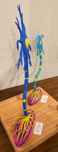
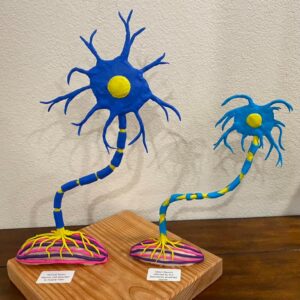
Amyotrophic Lateral Sclerosis 3D Image of a healthy motor neuron attached to a healthy muscle fiber and a diseased, dying motor neuron attached to an atrophied muscle fiber.

Amyotrophic Lateral Sclerosis 3D Image of a healthy motor neuron attached to a healthy muscle fiber and a diseased, dying motor neuron attached to an atrophied muscle fiber.
Good job! Next time be sure to include your name with your submission Tracy Ingraham!
Whoops I see it at the top, on the banner. Please casually disregard my blind mistake.
Tracy’s paper on Amyotrophic Lateral Sclerosis, or ALS, focuses on the pathology, effects, and potential causes and treatments of the disease as the medical and scientific community continues to learn more about this devastating condition. ALS is a progressive, degenerative disease that is caused by the deterioration and death of motor neurons, specifically in the spinal cord and brain. Every skeletal muscle fiber is innervated by a motor neuron at the neuromuscular junction and these muscle fibers contract when activated by excitation signals from the neuron. In healthy muscle tissue, upper motor neurons direct lower motor neurons to produce muscle movement, but with muscle tissue affected by ALS, upper and lower motor neurons fail, which causes the inability of muscles to contract as they should. At first, stiffness, muscle twitches, and spasms occur, and as the disease progresses and muscles contract less and less, they atrophy and eventually, paralysis occurs. ALS, (also known as Lou Gherig’s disease), is fatal. Death most often occurs as the result of respiratory paralysis. Treatment of ALS remains largely palliative, focusing mostly on making patients’ lives more comfortable and high-functioning. However, there are currently two medications that are approved for treating ALS, edaravone and riluzoe. These drugs have proven effective in the treatment of ALS. There are many studies that have been and are currently being conducted regarding ALS, but much more research is needed to determine potential genetic factors, environmental contributions, and treatments/cures.
For the physical, STEM portion of Tracy’s project, to accompany her paper, she built two 3D models of motor neurons and muscle fibers. One model is a healthy motor neuron attached to a healthy muscle fiber, and the second is a diseased, dying motor neuron attached to an atrophied muscle fiber. The second model obviously represents a motor neuron and muscle fiber suffering from Amyotrophic Lateral Sclerosis. I am unsure of the medium she used; maybe colored clay or painted clay. It is really pretty, which sounds weird, since it is motor neurons and muscle fibers, but it is. It highlights how Lou Gherig’s disease affects the motor neurons and interferes with muscle function. I think this project is really interesting, incredibly well researched, and informative. The model of the motor neurons and muscle fibers should be in a classroom somewhere because seeing it helps make it easier to understand what is going on inside our muscles at the microscopic level and makes identifying the structural points of a motor neuron much more clear. Great job, Tracy!
Thank you Toby!!!