Scleroderma

Scleroderma is an autoimmune connective tissue that causes inflammation in the skin, blood vessels and can damage other areas of the body. There are a number of conditions that can cause scleroderma, such as rheumatoid arthritis, lupus, or Sjogren’s syndrome. It commonly affects the skin, making it hard and tight, but it can also affect organs. In my research, I found little information about why scleroderma occurs; the common theories are that it results from autoimmunity, genetics, and environmental factors. Although it is rarely ever found when not associated with certain autoimmune diseases. Scleroderma can be mild to severe. A milder form of this disease affects the skin and blood vessels due to the abnormal growth of connective tissue. This is called localized scleroderma. Resulting in the immune system malfunctioning, causing the fibroblasts it contains to produce too much collagen in the lungs, kidneys, and skin. The immune system does this because it thinks the tissues are damaged and is trying to repair them with lymphocytes. The lymphocyte is a white blood cell that contains killer cells, T-cells, and B-cells; it attacks the tissues activating apoptosis, the death of cells. 

Connective tissue is made up of five significant cells: fibroblasts, mast cells, plasma cells, macrophages, adipocytes, and leukocytes. When the activated T-cells and the macrophages become infiltrated, the mast cells are degranulated in the skin. Directly correlating with the malfunctioning of connective tissue that begins to break down, thus causing it to thicken. Collagen takes up 65-80% of the dry weight of the tendons. When it starts to be overproduced, the tendons’ structure starts to change. The change in the tendons starts to affect the connective tissues, which then become inflamed, and the skin becomes tight and damaged. The connective tissue supports and protects other tissues and organs; because of this, people with scleroderma often have swollen blood vessels, rashes, and joint pain; in the worst cases, the organs can be affected.

Scleroderma affects women far more than men. These women are often between the ages of 30-50 and, more commonly, women of color. African-Americans develop scleroderma earlier than other races, and when they do develop scleroderma, it is often very severe and affects the organs, most commonly the lungs and heart, and can even cause lung disease. Scleroderma is separated into two groups, localized and systematic. Localized scleroderma appears either linear, where it is a thick scar-like line running along the body, most often on the face or legs, or morphea, which presents in patches on the skin, and in some cases, it appears as both. Systematic scleroderma can be a limited cutaneous scleroderma, which gradually affects the fingers, face, hands, lower arms, and legs below the knees. The other kind is diffuse cutaneous scleroderma, which spreads quickly, starting at the fingers and toes, then spreading to the upper arms and thighs. Diffuse cutaneous scleroderma is often the type associated with organ damage. The build-up of the connective tissue and collagen causes scleroderma to look like a rash-type scar, stretching and pulling on the skin.

References

Mayo Clinic. (2019). Scleroderma – Symptoms and causes. Mayo Clinic. https://www.mayoclinic.org/diseases-conditions/scleroderma/symptoms-causes/syc-20351952

Hou, W. (2011). Scleroderma. https://doi.org/10.1016/b978-0-443-06974-1.00014-2

Nancy Garrick, D. D. (2017, April 12). Scleroderma. National Institute of Arthritis and Musculoskeletal and Skin Diseases. https://www.niams.nih.gov/health-topics/scleroderma#:~:text=Scleroderma%20is%20an%20autoimmune%20connective

Takahashi, T., Asano, Y., Sugawara, K., Yamashita, T., Nakamura, K., Ryosuke Saigusa, Ichimura, Y., Toyama, T., Taniguchi, T., Kaname Akamata, Noda, S., Yoshizaki, A., Tsuruta, D., Trojanowska, M., & Sato, S. (2017). Epithelial Fli1 deficiency drives systemic autoimmunity and fibrosis: Possible roles in scleroderma214(4), 1129–1151. https://doi.org/10.1084/jem.20160247

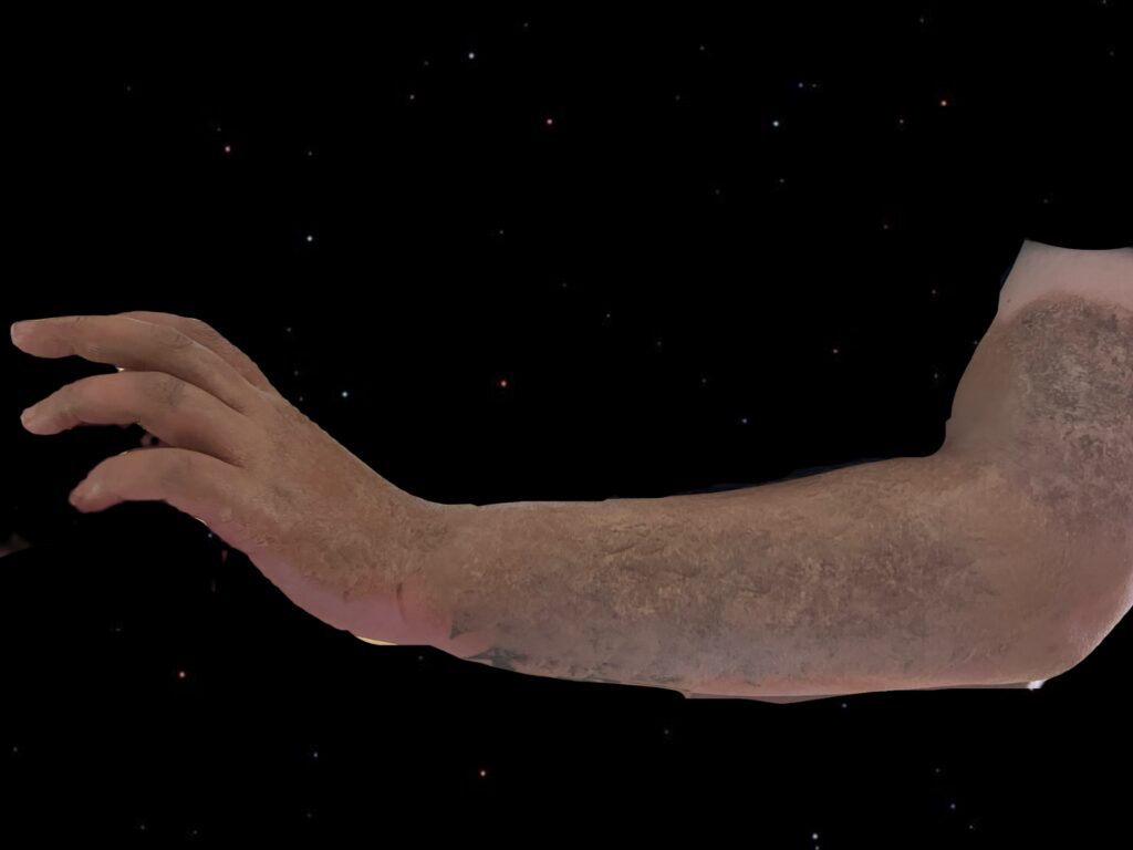
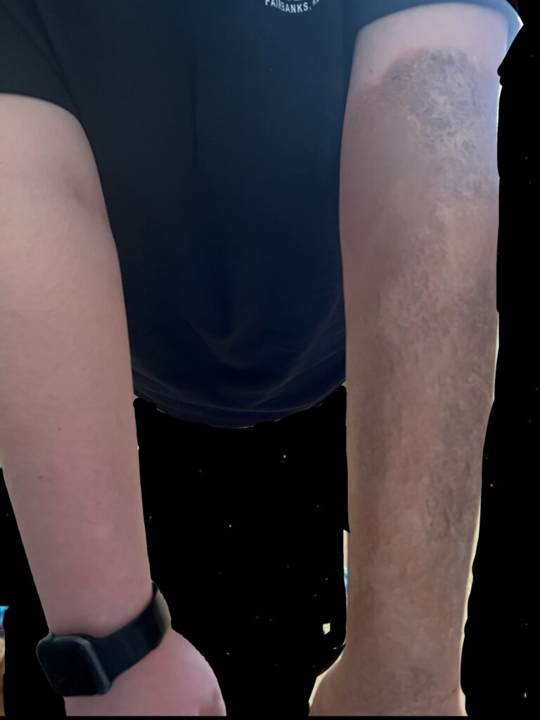
In my photos below, I decided to use makeup to show what scleroderma looks like on the surface of the skin. I had tried to make my hand appear swollen, but it did not turn out well, so I decided not to use those photos.

itemprop="discussionURL"One Comment

Burn Induced Cachexia

For My steam project I chose to research Cachexia in association with burn victims. As shown in the ink drawing Above. A man is in a burning room and is exposed to flames. He is burned and is depicted and skinny and frail, relating to the effects of Cachexia.

This work is meant to Identify the effect of cachexia/Hypermetabolism on the Skeletal Muscle and bone tissues in response to burning wounds. It is also meant to identify the aforementioned tissues and their functions.

Victims who suffer burns from stage 4 and up face a lot of issues when beginning the healing process. Along with the lymphatic system, respiratory system, and nervous system, major tissues that are badly affected are the Skeletal and muscle tissues. Both of these are fairly regenerative tissues but muscle tissue more so, and while they do undergo a healing process, they are not the same on a cellular level after being damaged by burns. These two tissues are the focus of burn induced cachexia and hypermetabolism, which cause dysfunction in muscle and bone tissues.

Bones(Osseous tissue) are present throughout the body, supporting other tissues. When burned in an accident they can face a multitude of issues, the first of which being bone loss or loss of bone mineral content(BMC). This burn injury results in a rise of proinflammatory cytokines which are as the name states “Proinflammatory” and do not promote healing. Through a chain of cellular interactions, these cytokines stimulate osteoclast activity. Therefore hastening bone resorption into the bloodstream. Other factors also play a role in the stimulation of osteoclast activity, and inhibition of osteoblasts. This means that bone resorption or degradation is increased while bone production is lowered. 

It is also notable that Parathyroid hormone(PTH) is much lower than what is required to regulate the calcium and phosphorus in the body in burn victims. Thus the calcium metabolism is no longer in good condition to regulate our calcium levels in a productive way.

Our Skeletal muscle is all over the body with origins and insertions along our bones. These tissues are in charge of voluntary movement, locomotion, and environmental manipulation. These contractile tissues are adversely affected by burns, and the key term to understanding burn induced muscle cachexia is hypermetabolism. Hypermetabolism is an increase in resting metabolic activity, in this case in response to the burn wound. The now hyperactive metabolism sets off an increase of stress regulators, such as catecholamines and glucocorticoids. These induce hypercatabolic responses, particularly proteolysis, which is the breakdown of proteins and peptides. This breakdown of proteins in the muscle, as well as other miscellaneous tissues within the body, become the fuel source for the hyperactive metabolism, and are used to heal the wound. 

This degradation of tissues due to a disturbed calcium metabolism, and a hyperactive metabolism weakens the body in order to heal burn wounds, and continues to be catabolic after the wound has healed, even continuing three years after the injury takes place. This is costly to the body and can actually bring harm to the body in the form of sepsis, organ failure, early stages of osteoporosis, and other medical complications. 

Burns and wounds. Burns and Wounds | Johns Hopkins Medicine. (n.d.). Retrieved November 8, 2022, from https://www.hopkinsmedicine.org/health/conditions-and-diseases/burns#:~:text=Second%2Ddegree%20burns%20involve%20the,may%20be%20swollen%20and%20painful.&text=Third%2Ddegree%20burns%20destroy%20the,bones%2C%20muscles%2C%20and%20tendons 

Cherney, K. (2019, June 4). 4th degree burn and other degrees: Classification, healing, more. Healthline. Retrieved November 8, 2022, from https://www.healthline.com/health/4th-degree-burn  

Klein, G. L. (2006, August 8). Burn-induced bone loss: Importance, mechanisms, and management. Journal of burns and wounds. Retrieved November 8, 2022, from https://www.ncbi.nlm.nih.gov/pmc/articles/PMC1687146/ 

Knuth, Carly M, et al. “Burn-Induced Hypermetabolism and Skeletal Muscle Dysfunction.” American Journal of Physiology. Cell Physiology, U.S. National Library of Medicine, 1 July 2021, https://www.ncbi.nlm.nih.gov/pmc/articles/PMC8321793/ 

CA;, Dinarello. “Proinflammatory Cytokines.” Chest, U.S. National Library of Medicine, Aug. 2000, https://pubmed.ncbi.nlm.nih.gov/10936147/  

itemprop="discussionURL"One Comment