
Osteogenesis imperfecta (OI) an inherited connective tissue disorder that mainly affects bones. (Martin et al., 2007) Causes bones to be fragile and have low bone mass. (Rauch et al., 2006) There are currently seven known types of OI. (Martin et al., 2007) Type I few fractures and deformities vertebral fractures are common and may lead to mild scoliosis. (Rauch et al., 2006.) Type II baby is born with short limbs chest is small and skull is soft newborn is often born with fractured bones lungs are not developed low birth weight life expectancy is very low. (John Hopkins Medicine.) Type III babies’ limbs are shorter ribs have fractures the head is abnormal size triangle face and deformed chest scoliosis difficulty swallowing and breathing. (John Hopkins Medicine) Type IV moderate bone deformities and variably short stature. (Rauch et al., 2008) Type V will have ossification of the interosseous membrane at the forearm and a predisposition to develop hypertrophic calluses. (Rauch et al., 2006) Type VI mineralization defect in the bones and hyperosteoidosis on bone biopsy. (Martin et al., 2007) Type VII mutation affecting a protein called CRTAP. (Rauch et al., 2006) Type I-IV have collagen mutation at different levels and Type VII, II, III is the recessive lethal. (Martin et al., 2007) In Type I the collagen is affected where the glycine residue is in one of the two genes encoding collagen type I x chains instead of having it at every third position there are three polypeptide chain two x1 and one x2 chain (COL1A1 and COL1A2). (Roschger et al., 2008) This is known as qualitative mutation. (Roschger et al., 2008) In children the severe forms of OI the bone matrix is more highly mineralized, leading to increased stiffness and hardness. (Roschger et al., 2008) The bone tissue in children is reduces bone mass impaired trabecular architecture, increased matrix mineralization density. (Roschger et al., 2008) Individuals that have Type I III and IV the osteoblasts produce half the amount of bone matrix. (Roschger et al., 2008) Type I is collagen mutation the quantitative is at 50% of normal procollagen chains are secreted. (Martin et al, 2007) Type II collagen mutation dominant structural mutations in COL1A1 and COL1A2. (Martin et al., 2007) How these effects the collagen is by the abnormal protein is formed leading to formation of disorganized collagen polymer and matrix disruption. (Martin et al., 2008) Type III collagen mutation dominant the structural mutations in COL1A1 and COL1A2. (Martin et al., 2008) How this affects the collagen is the abnormal protein is formed leading to formation of disorganized collagen polymer and matrix disruption. (Martin et al., 2008) Type IV collagen mutation dominant structural mutations in COL1A2 meaning there is an abnormal protein is formed, leading to formation of disorganized collagen polymer and matrix disruption. (Martin et al., 2008) OI will have blue sclera, dentinogenesis imperfecta, hyperlaxity of ligaments and skins, hearing impairment, and Wormian bones on the skull’s radiographs. (Rauch et al., 2006) Depending on the type of OI you have will determine what kind of treatment the child will be able to receive. Type III and IV are best treated with physiotherapy, rehabilitation, and orthopedic surgery. (Rauch et al., 2006) During these treatments physical activity program is encouraged to help prevent contractures and immobility induced bone loss. (Rauch et al., 2006) Orthoses is what is used to prevent the lower limbs in the earlier phases. (Rauch et al., 2006) There is also Bisphosphonate therapy is an antiresorptive agent that helps inhibit osteoclast function. (Rauch et al., 2006) The whole purpose of this treatment is to decrease the activity in the bone resorbing system in hopes it will compensate for the weakness of the bone- forming cells. (Rauch et al., 2006) Another treatment that helped with bone pain is cyclical intravenous pamidronate and are given 1 to every 4 months and helped with an increase of well-being, and a rapid rise in vertebral bone mineral mass. (Rauch et al., 2006) Growth Hormone is another treatment for OI this could accelerate short term height velocity, the bone turnover increases but the calcium retention is unchanged. (Rauch et al., 2006)
The art piece shows a health bone compared to one that has osteogenesis imperfecta, normal triple helix and one that has OI, what a wormain skull looks like and an abnormal skull. The objectve covered was to explain how bone development is hormonally regulated. This essay explained how OI affects children’s bones development. Depending on the type of osteogenesis imperfecta the child has, will determine the effect on the bone’s collagen.
Sources
Martin, E., Shapiro, J.R. Osteogenesis imperfecta: Epidemiology and pathophysiology. Curr Osteoporos Rep 5, 91–97 (2007). https://doi.org/10.1007/s11914-007-0023-z
Osteogenesis imperfecta. Johns Hopkins Medicine. (2022, July 19). https://www.hopkinsmedicine.org/health/conditions-and-diseases/osteogenesis-imperfecta
Rauch, F., Glorieux, F.H. Treatment of children with osteogenesis imperfecta. Curr Osteoporos Rep 4, 159–164 (2006). https://doi.org/10.1007/s11914-996-0025-2
Roschger, P., Fratzl-Zelman, N., Misof, B.M. et al. Evidence that Abnormal High Bone Mineralization in Growing Children with Osteogenesis Imperfecta is not Associated with Specific Collagen Mutations. Calcif Tissue Int 82, 263–270 (2008). https://doi.org/10.1007/s00223-008-9113-x
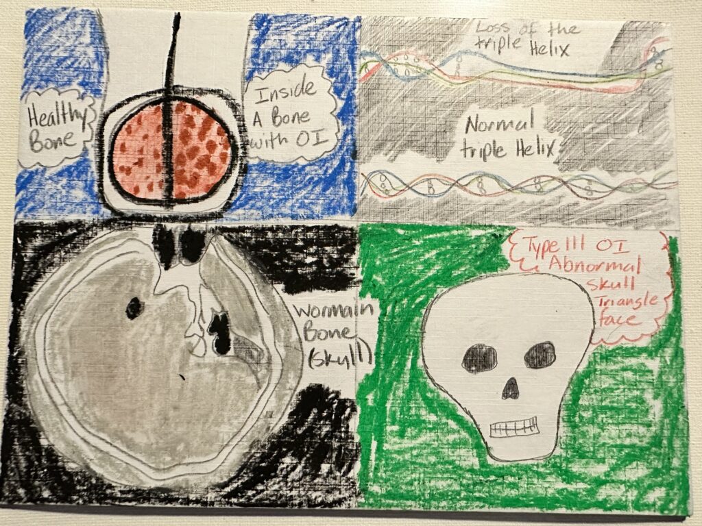
The art piece shows a healthy bone, a normal triple helix and a Wormian skull compared to one that has osteogenesis imperfecta (OI) or abnormal skull. OI is an inherited connective tissue disorder that affects bones, collagen, and the proteins that provide structure and strength to our tissues. There are seven types of OI. Each with different severities and symptoms. Some of the common features of OI are low bone mass, fragile and easily broken bones, blue sclera, dental problems, hearing loss, and loose skin and joints. The cause of OI is a mutation in the type I collagen that is encoded by the COL1A1 gene, type 1 collagen makes up most of the body’s total collagen and the most abundant in the bones. This mutation affects the quantity or quality of collagen and leads to abnormal bone formation and mineralization. A diagnosis of OI is based on family history, and genetic testing. Depending on the type of OI the person has will determine what kind of treatment they will receive. The main treatments are physiotherapy which type III and IV are best treated with this, it is a physical activity program to help prevent contractures and immobility induced by the bone loss. Another treatment option is orthopedic surgery, or bisphosphonate therapy, which is a medication that inhibits osteoclast formation. The whole purpose of this treatment is to decrease the activity in the osteoclast in hopes it will compensate for the weak osteoblast. The treatment of OI is directed to prevent fractures, improve mobility, reduce pain and improve quality of life.