 Posted by
November 20,2025
Craniotomy Surgery
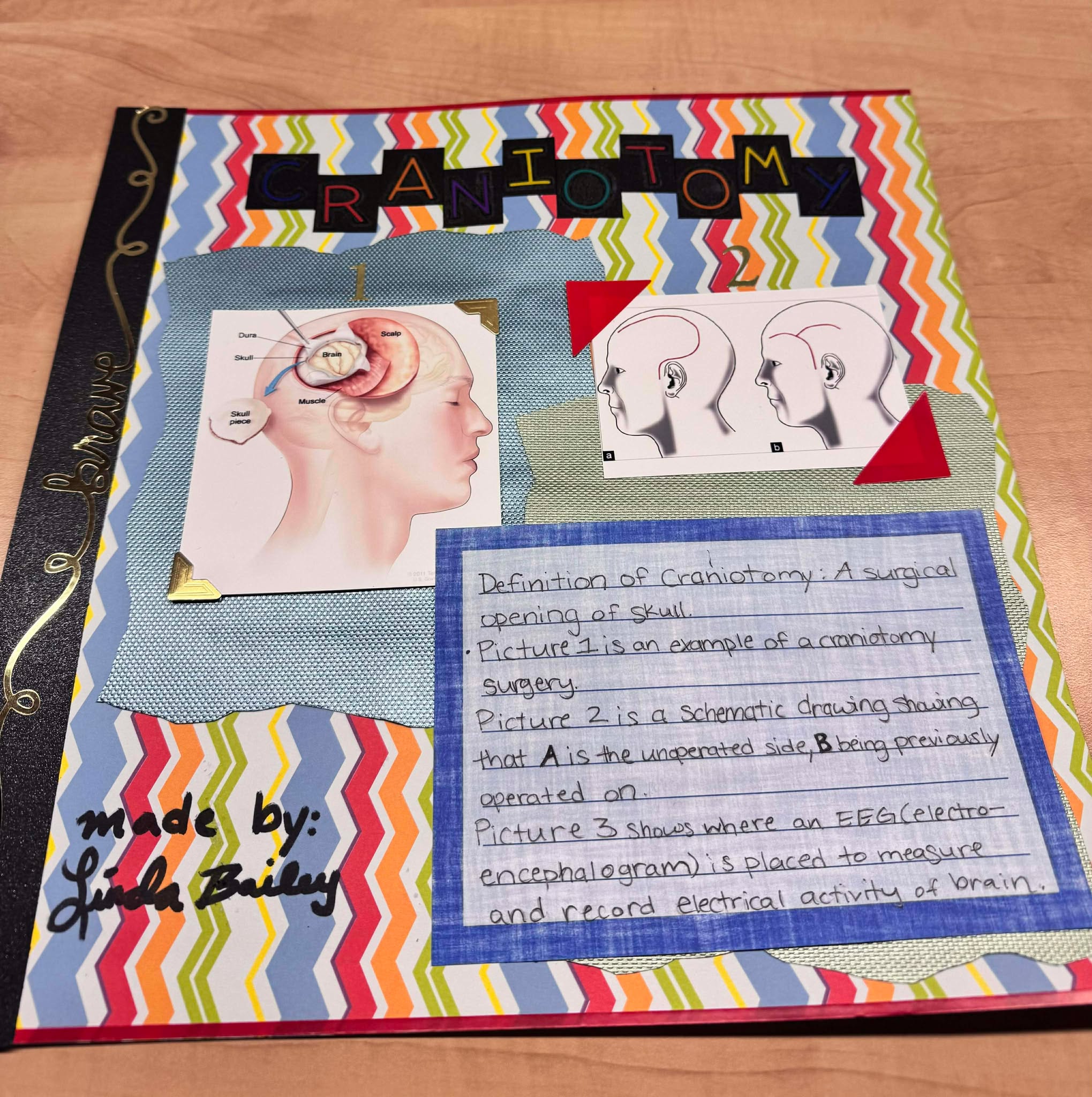
The definition of craniotomy is a surgical opening of the skull. The topic I have decided to focus on for my steam project is about craniotomy surgeries. The time of 5000-8000 BC was the first recorded craniotomy in history. The purpose of early craniotomies in 8000-5000 B is not yet fully understood. Some believe it would help therapeutically in response to trauma (Rao et al., 2020). Reminds me of lobotomies. Some even used craniotomies for reasons they believed related to magic, religious rituals or initiation practice (Sperati, 2007). Crazy! How vast the improvements we have seen and documented overtime. Some believe craniotomies have been practiced longer than any other procedures, making it significant. Well, if you’re determining that by evidence, it goes all the way back to the neolithic period which is in 8000-5000 BC. An example of discovering more about the brain overtime is discovering that the foreman was linked to trauma until 1783. Since then, we are able to identify parts of the brain and their functions more accurately. Still to this day we are advancing.
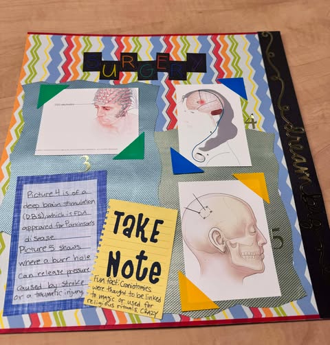
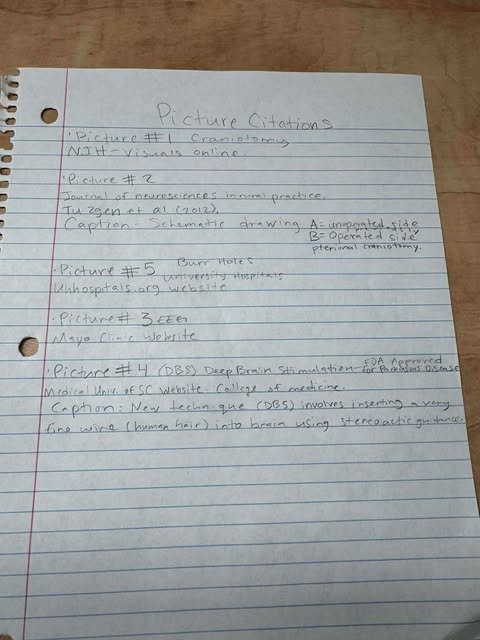
Later, craniotomies were used for treatment purposes as little as headaches. It was also used for removing tumors, clip or repair aneurysms, malformations, and tears or fractures in the dura matter. In relevance, my older brother had a blood clot removed from a leaking blood vessel. In the pictures provided in my steam project, I have a (EEG) electroencephalographic recordings within skull to diagnose epilepsy. An implantation simulator device that was inserted to treat movement disorders like Parkinson’s. Burr holes (see picture) can also relieve pressure within brain. Removing damaged or swollen areas of brain caused by stroke or traumatic injury helps relieve pressure.
The objective I will be covering is the stages of bone development and repair, in the skull. The repair bone potential, including inflammatory and Schwann cells, continues throughout adulthood. Hence the saying, your frontal cortex isn’t fully developed until you’re twenty-four. Repair continues until tissue is repaired back to its homeostatic functional state. Bone forming osteoblasts and bone-resorbing osteoclasts are mostly responsible for bone repair according to studies (Salhotra et al., 2020).
I was there for my brother when he had a craniotomy surgery, until he recovered to a more stable state. This type of surgery has been around for many years, evolved within time. The evolution of medical imaging has improved rates significantly. My brother had pressure built up in his skull so they had to open up a part to release pressure. While the skull was off, a metal plate was placed. Until my brother was physically ready, they put his skull back in its original place. This was crazy to witness first hand, seeing half my brother’s skull missing.
Citations:
(Rao et al., 2020).
Rao, D., Le, R. T., Fiester, P., Patel, J., & Rahmathulla, G. (2020). An Illustrative Review of Common Modern Craniotomies. Journal of clinical imaging science, 10, 81. https://doi.org/10.25259/JCIS_176_2020Links to an external site.
(Sperati, 2007)
Sperati G. (2007). Craniotomy through the ages. Acta otorhinolaryngologica Italica : organo ufficiale della Societa italiana di otorinolaringologia e chirurgia cervico-facciale, 27(3), 151–156.
Salhotra, A., Shah, H. N., Levi, B., & Longaker, M. T. (2020). Mechanisms of bone development and repair. Nature reviews. Molecular cell biology, 21(11), 696–711. https://doi.org/10.1038/s41580-020-00279-w
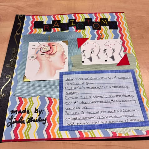
Linda Bailey made her STEAM project about craniotomy surgeries. Craniotomies are surgical openings of the skull. The course objective covered by her subject is the stages of bone development and repair, applied to the skull in this case. She explained the history of craniotomies and why they were used during these different times. The first recorded craniotomy took place in 8000-5000 BC. The reason why people were using craniotomy back then is not really clear. They thought it would help with trauma or even reasons related to magic. The purpose of this surgical procedure is now more specific and scientifically supported. It is used for removing tumors, malformations, fractures, or even blood clots (in the case of her brother). She gave some more details about her brother and how the procedure took place and the rehabilitation process.
She included some examples of the procedure, used to diagnose different disorders: an electroencephalogram, to diagnose epilepsy; Burr holes to relieve pressure in the brain; deep brain stimulation, to treat Parkinson’s. She illustrated her project with pictures and some explanation for each picture.