250-word abstract: Wolff-Parkinson-White (WPW) syndrome is a heart rhythm disorder that happens when an extra electrical pathway in the heart interferes with the heart’s normal conduction system. In a normal sinus rhythm, electrical impulses originate in the sinoatrial (SA) node and travel through the atria, are briefly delayed at the atrioventricular (AV) node, and then are transmitted to the ventricles. This delay is necessary for the proper filling of the ventricles and coordinated contraction. In WPW, the electrical impulses go around the AV node via the bundle of Kent, an accessory pathway. This causes the ventricles to fire early, and the timing is off.
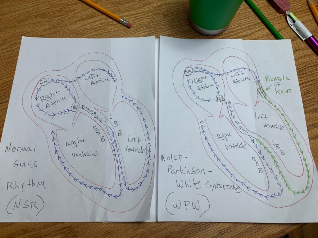
The STEAM project compares the normal sinus rhythm versus WPW with two heart diagrams. In the normal diagram, blue arrows follow the correct conduction pathway through the AV node. In the WPW diagram, the Bundle of Kent is shown by additional green arrows on the left of the heart. These arrows show the electrical signal going around the AV node, causing faster conduction and upsetting the normal sequence of contraction.
WPW is diagnosed on an electrocardiogram (EKG) by three important findings: a shortened PR interval, a delta wave at the beginning of the QRS complex, and a widened QRS complex. These features represent early activation of the ventricles. This condition develops in the fetal heart due to a defect in the annulus fibrosus, which acts as an electrical barrier.
If not correctly identified, WPW may lead to serious problems such as atrial fibrillation or ventricular fibrillation. Radiofrequency catheter ablation is a highly effective treatment that can eliminate the accessory pathway and often leads to a long-term cure.
 Posted by
250-word abstract: Wolff-Parkinson-White (WPW) syndrome is a heart rhythm disorder that happens when an extra electrical pathway in the heart interferes with the heart’s normal conduction system. In a normal sinus rhythm, electrical impulses originate in the sinoatrial (SA) node and travel through the atria, are briefly delayed at the atrioventricular (AV) node, and then are transmitted to the ventricles. This delay is necessary for the proper filling of the ventricles and coordinated contraction. In WPW, the electrical impulses go around the AV node via the bundle of Kent, an accessory pathway. This causes the ventricles to fire early, and the timing is off.
The STEAM project compares the normal sinus rhythm versus WPW with two heart diagrams. In the normal diagram, blue arrows follow the correct conduction pathway through the AV node. In the WPW diagram, the Bundle of Kent is shown by additional green arrows on the left of the heart. These arrows show the electrical signal going around the AV node, causing faster conduction and upsetting the normal sequence of contraction.
WPW is diagnosed on an electrocardiogram (EKG) by three important findings: a shortened PR interval, a delta wave at the beginning of the QRS complex, and a widened QRS complex. These features represent early activation of the ventricles. This condition develops in the fetal heart due to a defect in the annulus fibrosus, which acts as an electrical barrier.
If not correctly identified, WPW may lead to serious problems such as atrial fibrillation or ventricular fibrillation. Radiofrequency catheter ablation is a highly effective treatment that can eliminate the accessory pathway and often leads to a long-term cure.