 Posted by
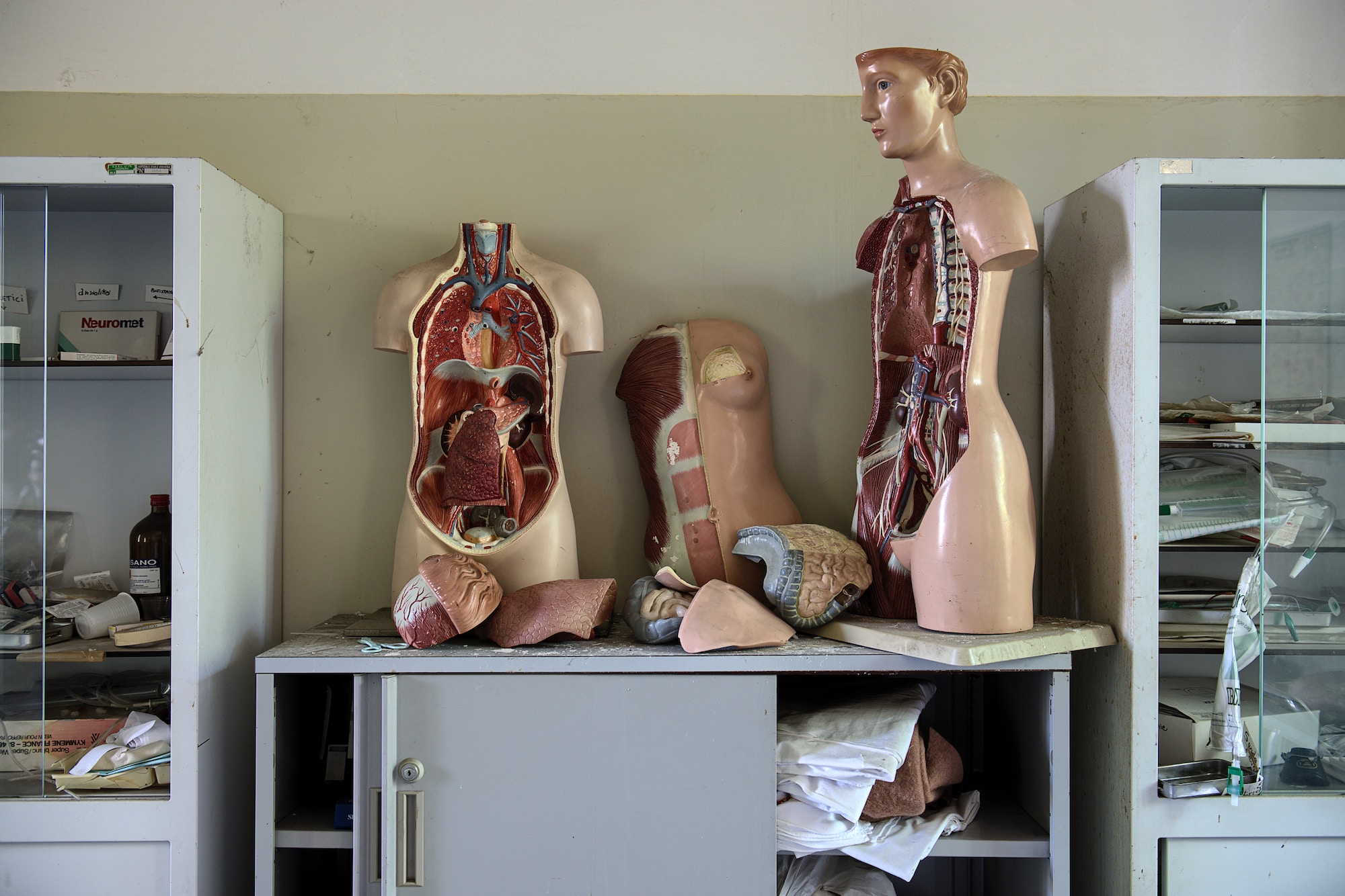
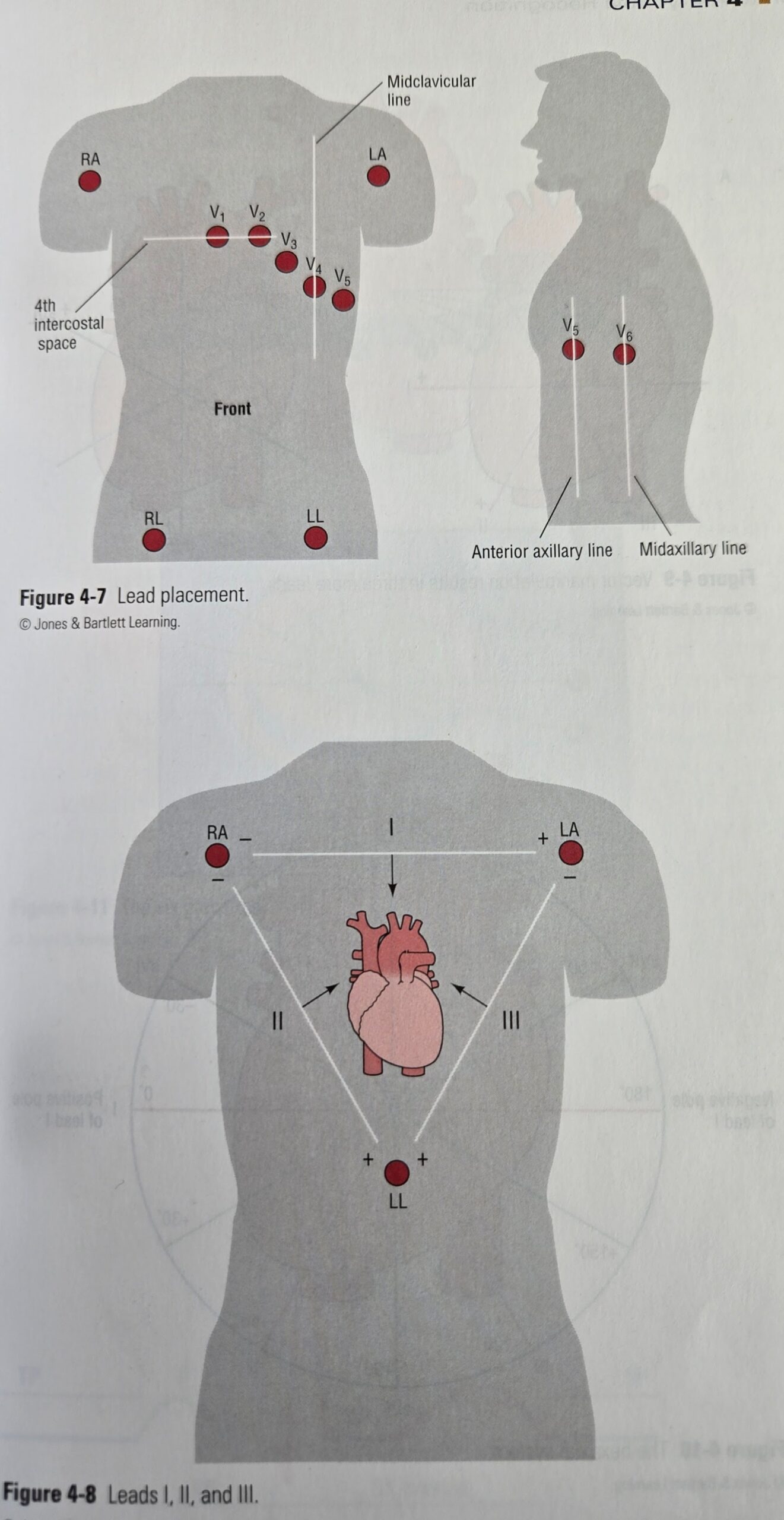
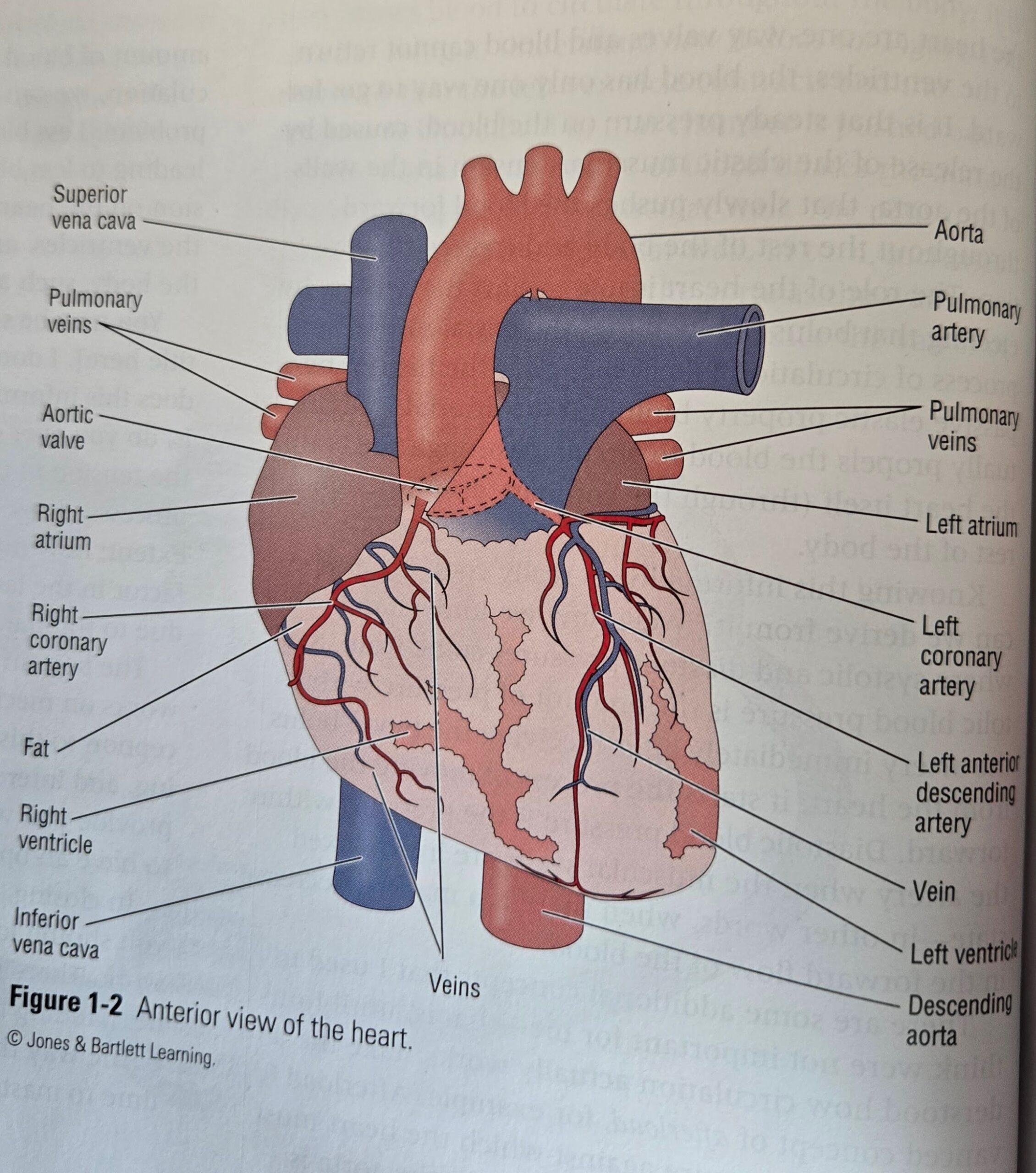
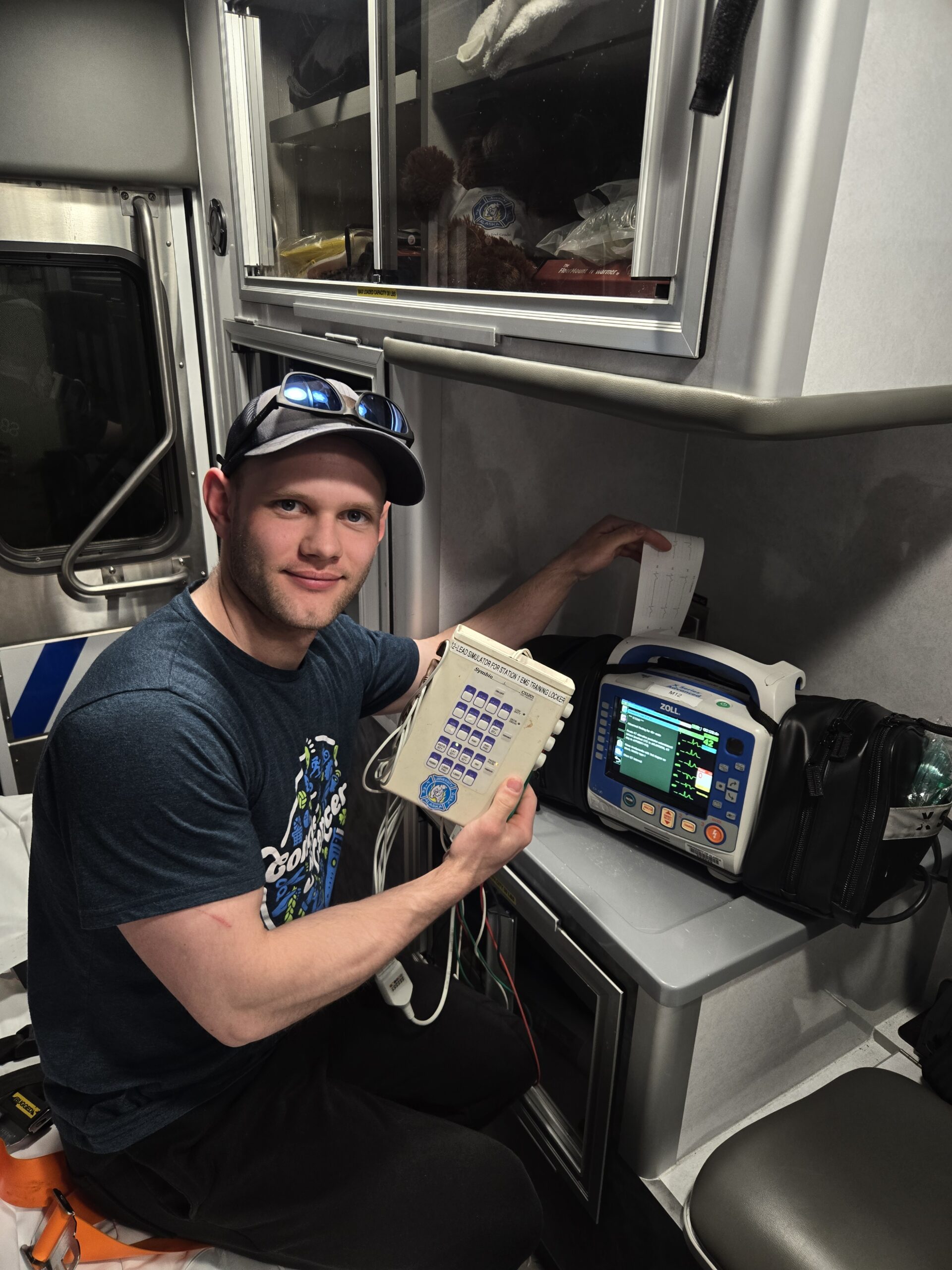
I have included two photos of 12-lead ECG rhythm strips that include three different STEMIs. I showed you how to identify a STEMI in the top two rhythms and left the bottom one for you figure out. I have also attached two helpful photos that show you where the electrodes are placed on a person as well as the location of the coronary arteries of the heart. Both of those those images were taken from the textbook Arrhythmia Recognition The Art of Interpretation 2nd edition by Tomas B. Garcia MD and Daniel J. Garcia, published by Jones & Bartlett Learning. The last photo is proof that I printed out the rhythms with what is called a “chicken heart.” It has many different rhythms to choose from and is used as a training device to learn how to interpret 12-leads.
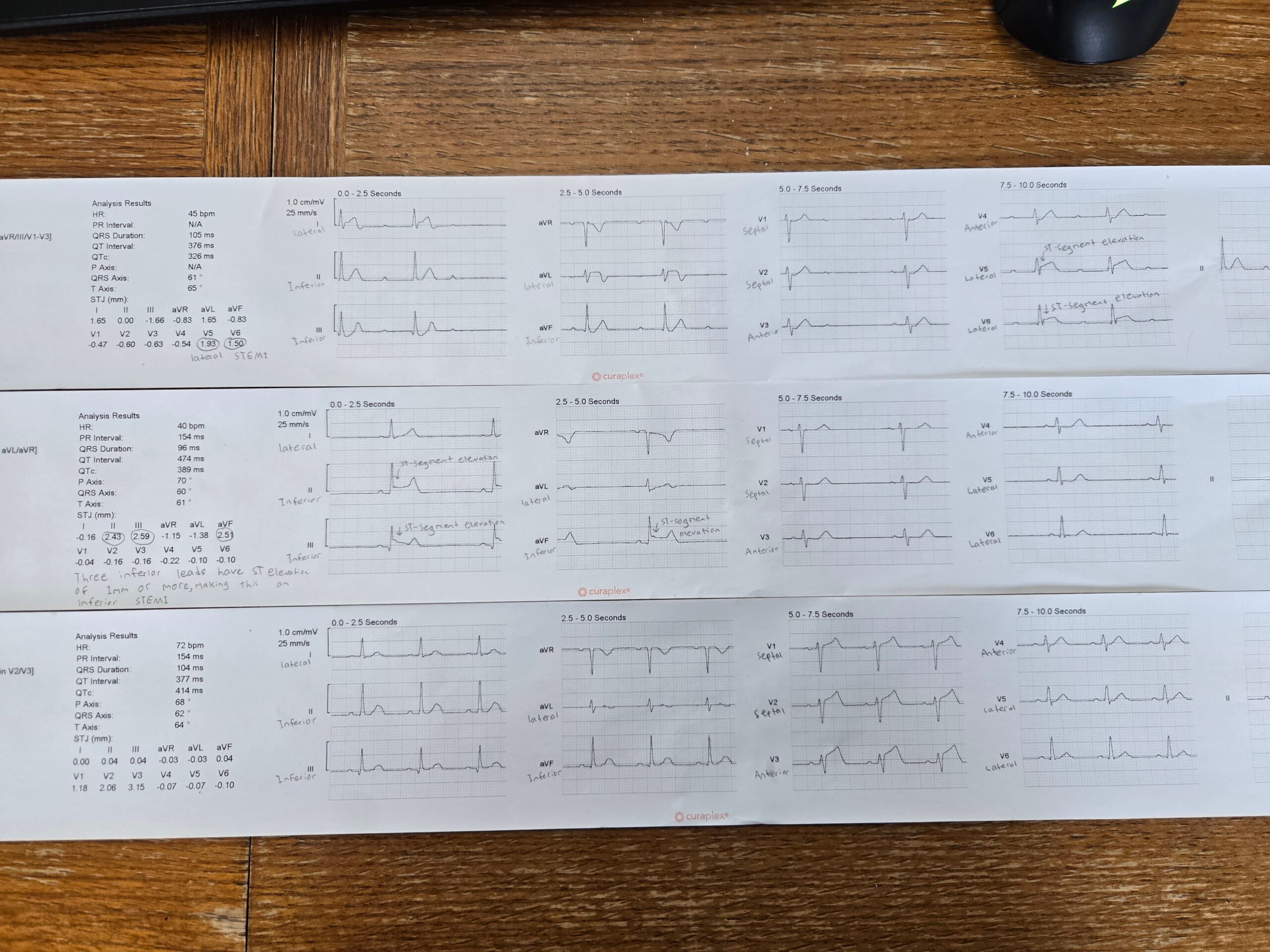
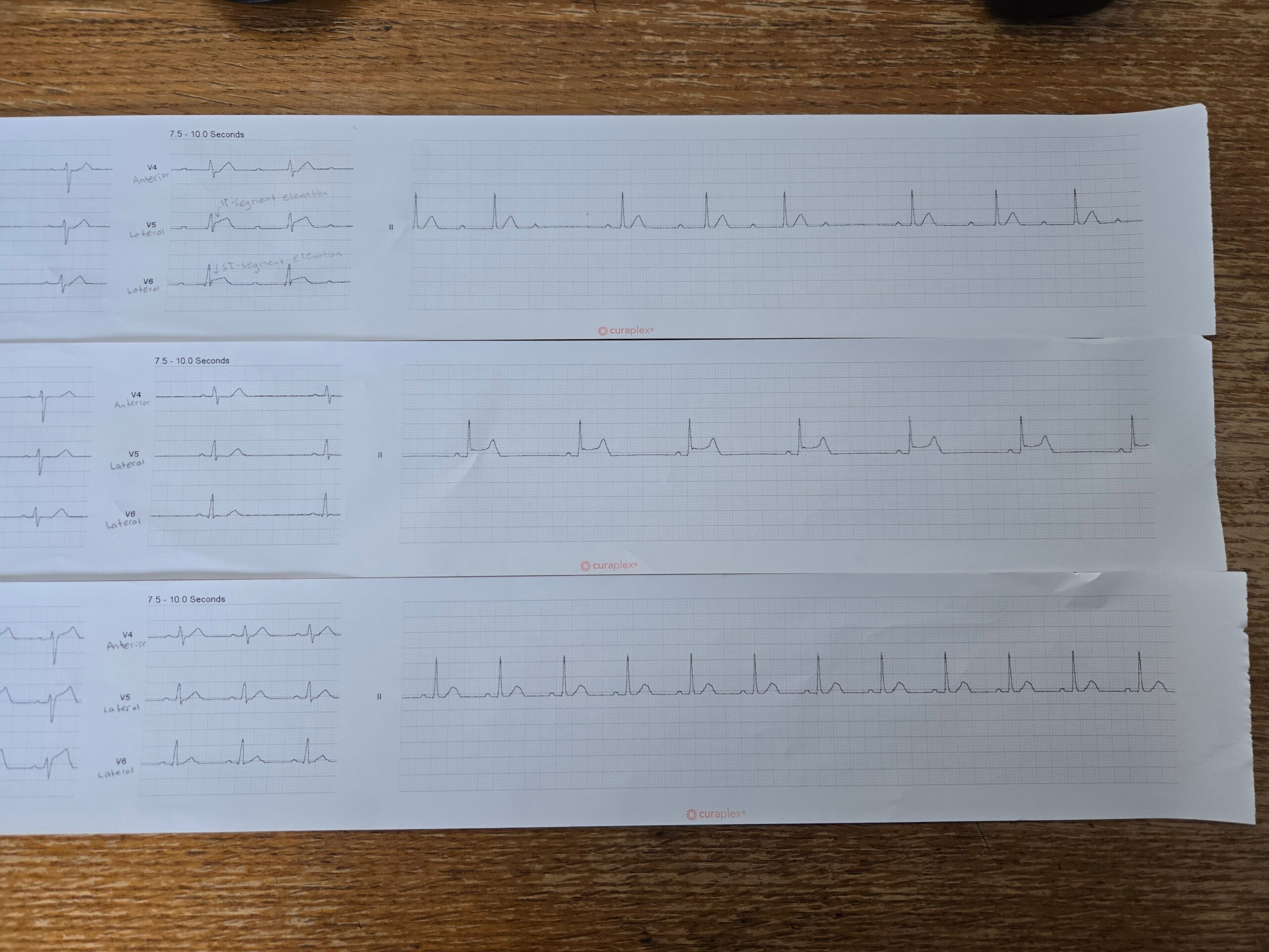
ST-segment Myocardial Infarction (STEMI) is a full blockage of a coronary artery. The primary way of identifying it is through 12 lead ECG. It is seen when two or more leads have a higher than 1 mm spike. Those spikes in the ECG are triggered by the repolarization and depolarization of different parts of the heart. Utilizing that typically predictable cycle, it allows for EMS/EMT and other health professionals to identify quickly, and step in to treat the blockage. Treatment depends on if the blockage is in the left or right coronary artery or the anterior descending artery. However, possible options for treatment include painkillers, oxygen, aspirin, nitroglycerin and close monitoring.
ST-segment Myocardial Infarctions (STEMI) are caused by a complete blockage of a coronary artery. Because STEMIs are a life threatening a time-sensitive cardiac emergency, assessment and identification of STEMIs are crucial in both prehospital and hospital settings. The primary diagnostic tool used to identify STEMIs is a 12-lead ECG, which relies upon electrodes placed in areas on the chest and limbs to assess the heart’s electrical activity. There are four limb lead stickers that include bilateral arms and legs. There are also six precordial leads that read different parts of the heart. Ultimately, this test produces a cardiac rhythm strip that can be interpreted. Early identification and intervention with reperfusion therapy can lead to significantly improved patient outcomes by restoring coronary blood flow and limiting myocardial damage.
After reviewing your project on 12-lead ECG interpretation and STEMI identification, I learned a great deal from how you broke everything down. You did a very good job showing how ST-segment elevation in certain leads points to different areas of the heart, such as the inferior, anterior, and lateral regions. That helped me understand more clearly how to recognize patterns quickly instead of focusing only on individual leads. What stood out to me most was how you linked the ECG changes to the coronary arteries. Seeing how a blockage in a specific artery directly affects certain leads made it much easier to grasp why those changes occur. Your use of labeled rhythm strips and clear examples made the material easier to follow and apply in practice. The project helped me feel more confident in identifying STEMIs and understanding what is actually happening in the heart during a myocardial infarction. It also reminded me how important it is to pay close attention to detail when reading a 12-lead ECG, especially in emergency situations where time is critical. Overall, I gained a lot from your work, and it gave me a stronger understanding of how to interpret ECGs more accurately.