 Posted by
Growing pains when it comes to bones are not just reserved for teenagers or kids. Humans experience constant renewal and remodeling of their bone structure from day to day. Bone remodeling is a process that takes multiple cells to complete, such as osteoclasts. Osteoclasts originate from hematopoietic stem cells found in bone marrow. These cells are multinucleated and resorb bone tissue to allow for bone growth (Xu & Teitelbaum, 2013). Additionally, osteoclasts have specialized cytoskeletons. This cytoskeleton allows for the osteoclast to polarize on the bone and break down the mineralized structure quickly (Teitelbaum, 2007).
Every step of the process is important to the outcome, even the creation of osteoclasts. Osteoclasts are created through a process named osteoclastogenesis. In this process, macrophage colony-stimulating factor (M-CSF) and RANKL send signals to monocytes called essential differentiation signals, which trigger fusion and activation for osteoclasts (Teitelbaum, 2007). This is the main reason why osteoclasts have multiple nuclei. Once created, osteoclasts get to work on remodeling the bone, but they must tightly attach to the bone to do so. Therefore, they create two separate formations. One of these formations is the sealing zone. Integrins attach themselves to both the osteoclasts and the bone. This creates an area with just the cell and the bone, called the resorption lacuna, and is surrounded by actin filaments (Teitelbaum, 2007). This allows for the second formation to continue. This creates the ruffled border that forms on the side of the osteoclast connected to the bone (Xu & Teitelbaum, 2013). The ruffled border increases surface area so the cell can release more enzymes and acids (Teitelbaum, 2007).
After the formations are created, the osteoclast finally attempts to resorb the defective bone. Resorption begins with the osteoclasts dissolving minerals within the bone formations. The cells distribute large amounts of hydrogen ions into the lacuna using vacuolar H-ATPases. Due to this sequence, the lacuna becomes acidic to dissolve hydroxyapatite, the mineral responsible for the bone’s density, and phosphate ions enter the bloodstream (Teitelbaum, 2007). To break the remaining organic material left behind, cathepsin K is released by the osteoclasts. Finally, the leftover material is degraded and pulled into the cell to be kept on the opposite side from the bone (Xu & Teitelbaum, 2013). Once the osteoclasts have removed all defective pieces of bone, the osteoblasts can reform the bone into a stronger formation.
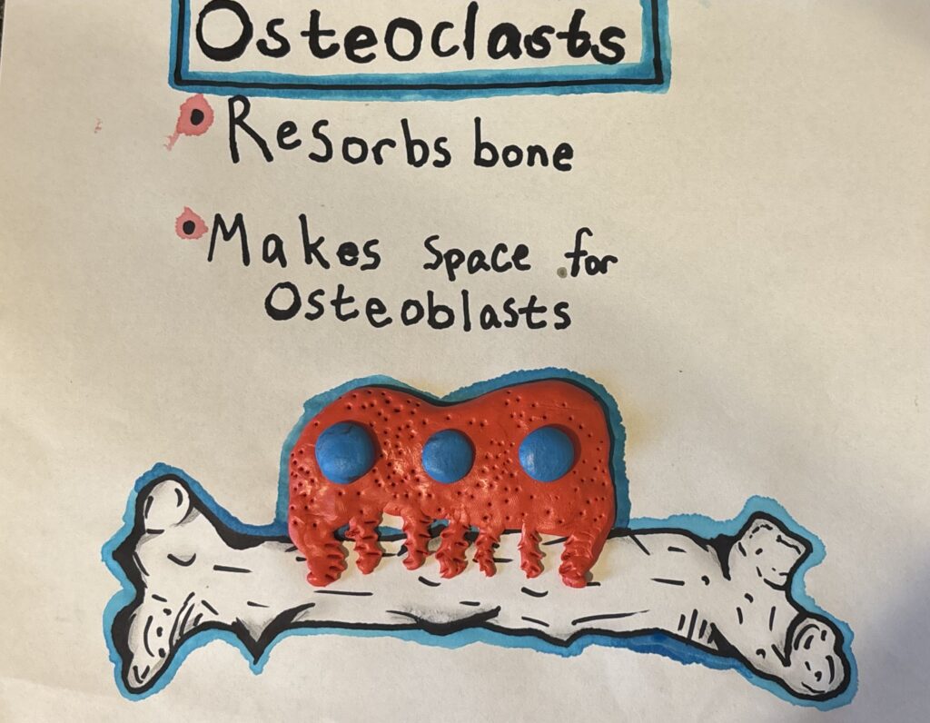
My STEAM project reflects this concept. I aimed to learn how osteoclasts function in comparison to the other types of bone cells. First, I drew the best bone I could manage (I’m no artist, but it’s recognizable at least). I then created a model of an osteoclast to attach to said bone. I referenced an image found in Teitelbaum’s article and created the model based on when osteoclasts deliver H-ATPases to the bone to distribute hydrogen ions. Overall, I learned a great deal from this project, specifically regarding how important osteoclasts are in regards to the upkeep of bones. It allowed me to work with my hands in a way that is usually not possible when studying cells.
References
Teitelbaum, S. L. (2007). Osteoclasts: What Do They Do and How Do They Do It? The American Journal of Pathology, 170(2), 427–435. https://doi.org/10.2353/ajpath.2007.060834
Xu, F., & Teitelbaum, S. L. (2013). Osteoclasts: New Insights. Bone Research, 1(1), 11–26. https://doi.org/10.4248/br201301003