 Posted by
For my STEAM project, I explored disuse atrophy in coma patients. I chose this topic because it beautifully connects basic muscle biology with real-world clinical challenges in critical care. When patients lose voluntary movement—especially if they are unconscious—their muscles can shrink rapidly, weaken, and lose their structure due to lack of stimulation. Understanding how normal muscle contraction works, why immobility speeds up muscle breakdown, and how healthcare providers try to slow this process helps us appreciate how sensitive the body is to inactivity. This project also highlights where physiology, pathology, and medical technology come together.
A typical muscle contraction starts at the neuromuscular junction, where a motor neuron releases acetylcholine, triggering an action potential in the muscle fiber. This electrical signal spreads along the sarcolemma and T-tubules, prompting the sarcoplasmic reticulum to release calcium ions into the cytoplasm. Calcium binds to troponin, shifting tropomyosin and revealing active sites on actin filaments. Myosin heads attach to actin and, through ATP-driven cross-bridge cycling, shorten the sarcomeres. This is called excitation–contraction coupling, and it depends on continuous neural signals and mechanical load. When these signals stop, like in long-term unconsciousness, muscle tissue no longer gets the cues needed to maintain protein balance and structure.
Research shows disuse atrophy causes widespread molecular changes in skeletal muscle. Miura et al. (2017) used proteomic and transcriptomic analyses to show that inactivity significantly decreases the expression of key structural proteins and mitochondrial enzymes, while increasing pathways for protein degradation. Their study highlights the upregulation of the ubiquitin-proteasome system, which is a main mechanism for muscle protein breakdown during disuse. Barreiro et al. (2018) added that disuse also reduces protein synthesis, leading to oxidative stress, mitochondrial problems, and inflammation—all of which speed up muscle loss. In coma patients, these changes tend to happen faster due to additional stressors like illness, immobility, inflammation, and poor nutrition.
Muscle atrophy in ICU patients isn’t just about appearance; it has serious consequences for recovery, mobility, and long-term health. Losing muscle mass increases the risk of infections, pressure ulcers, and dependence on ventilators. Even after waking up, many patients need months of rehab to regain strength and function. That’s why preventing or slowing muscle loss is so important in intensive care.
A promising method is electrical muscle stimulation (EMS). Hirose et al. (2013) found EMS helped reduce muscle atrophy in ICU patients with impaired consciousness compared to those who didn’t receive stimulation. EMS delivers controlled electrical impulses that cause muscles to contract, mimicking natural voluntary movements. While it can’t fully replace normal activity, EMS helps keep muscles active, maintains circulation, and slows deterioration during periods of immobilization. This technology shows how medical devices can help fill in for lost physiological functions.
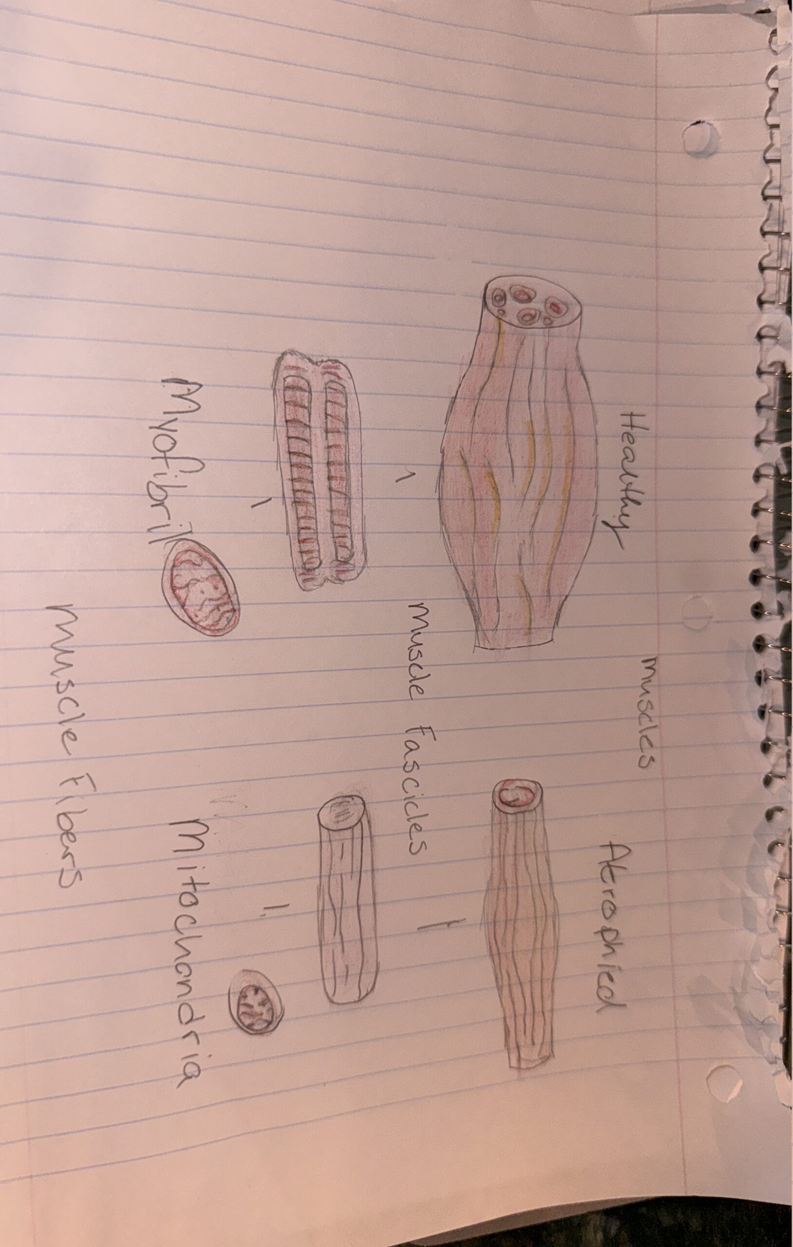
For my artistic part, I made a detailed illustration comparing healthy skeletal muscle fibers to atrophied ones. Healthy fibers are thick with dense myofibrils and many mitochondria, showing strong, active muscles with high metabolic capacity. The atrophied fibers are thinner, less organized, and have a smaller cross-sectional area. Insets show cellular differences, including fewer myofibrils and lower mitochondrial density, matching research findings. I also included a small diagram showing how electrical stimulation triggers muscle contraction. Creating this illustration helped me see and communicate how muscle structure changes a lot without neural and mechanical signals.
References
Barreiro, E., et al. (2018). Models of disuse muscle atrophy: therapeutic implications. Journal of Cachexia, Sarcopenia and Muscle.
https://pmc.ncbi.nlm.nih.gov/articles/PMC5799135/
Hirose, T., Shiozaki, T., Shimizu, K., Mouri, T., Noguchi, K., Ohnishi, M., & Shimazu, T. (2013). The effect of electrical muscle stimulation on the prevention of disuse muscle atrophy in patients with consciousness disturbance in the intensive care unit. Journal of Critical Care, 28(4), 536.e1–536.e7.
https://www.sciencedirect.com/science/article/abs/pii/S0883944113000531
Miura, T., et al. (2017). Disuse atrophy of skeletal muscle: proteomic and transcriptomic mechanisms. Journal of Applied Physiology.
https://journals.physiology.org/doi/full/10.1152/japplphysiol.00985.2016