 Posted by
Contracture is a prevalent condition stemming from many other medical conditions and is defined as a permanent or persistent shortening of a muscle, tendon, joint capsule, or other soft tissue. I will be focusing on muscle contracture for the purpose of this paper. The course objective I will be examining and associating with my topic is “ describe how a muscle contraction is induced”. My project will represent a normal functioning elbow and an elbow physically altered from contracture using pvc pipe and balloons.
A muscle contraction is the process in which a muscle generates tension and shortens or maintains tension without shortening in response to a nerve impulse, allowing movement of our bodies. During a muscle contraction nerves communicate an action potential to the muscle through a neuromuscular junction. Here ACH is released into the synaptic cleft. This ACH binds to receptors on the Sarcolemma, causing depolarization. In the beginning of a muscle contraction excitation contraction coupling occurs as the action potential now in the muscle tells the sarcoplasmic reticulum to release calcium ions into the cytoplasm. This causes actin and myosin to interact and start the Crossbridge cycle, a sequence of enzymatic reactions. As the calcium binds to troponin, tropomyosin moves exposing the binding sites on the Actin filaments. The myosin heads attached to the Actin binding sites and cross bridges are formed. As ATP is hydrolyzed to ADP and Pi or inorganic phosphate this release of Pi drive the sliding of the filaments or the “power stroke” that produces force and contraction. This “power stroke” occurs as the myosin heads pull on the actin and filaments shortening the Sarcomere and a contraction occurs. Next the ACH is broken down stopping further depolarization, calcium ions are pumped back into the Sarcoplasmic reticulum and actin binding sites are recovered, and the muscle returns to its resting state.
Contractures are debilitating physical changes involving shortening and stiffening of tissues in the body. While common, they are almost always secondary to another condition. Common causes of contracture include neurological causes, muscular causes, connective tissue and joint causes, environmental causes, and some metabolic causes. I will be focusing on the neurological causes of contracture including spasticity from upper motor neuron lesions and peripheral nerve injury. Contracture is an involved multi factored process, and it is not directly caused by spasticity rather it is a gradual physical change in the muscle tissue. Muscle stiffness can be classified as active and passive where active is due to contraction of muscle fibers and passive due to changes in mechanical properties. So while spasticity is not the direct cause of contracture, it is the underlying cause of the eventual progression of loss of range of motion from muscle damage and atrophy in some people. Conditions that contribute to neurological damage leading to spasticity and nerve damage include stroke, cerebral palsy, multiple sclerosis, spinal cord injuries and traumatic brain injuries to name a few. Spasticity affects millions of people worldwide, approximately 80 to 90% with multiple sclerosis 70 to 80% of individuals with cerebral palsy 60 to 70% with spinal cord injury 30 to 40% with Parisis due to stroke and 30 to 50% with traumatic brain injury (Raghavan, 2025). This spasticity causes overactive neural signals, causing muscles to remain in a shortened state. Peripheral nerve injury is another cause affecting movement after UMN damage. Spasticity occurs in the very beginning of muscle contraction and interferes with the signals being sent to the muscle and does not actually interrupt the actual steps in muscle contraction. When normal brain control is altered and lower motor neurons become over excitable spasticity is the result. This causes hyperactive stretch reflexes reacting too quickly and strongly, causing excessive contraction or jerking when limbs move and velocity dependent stiffness, causing increased stiffness with movement. Spasticity leads to chronic muscle over activity. Overtime this causes increased resting muscle tension, reduced range of motion and muscles that will no longer reach full length. Actin and myosin cross bridging still occurs, just at a much more limited range of motion. This extended period of shortening is the first physical driver of contracture formation with constant spasticity leading to long-term immobility. The muscle starts to undergo changes like the loss of Sarcomeres, when this happens, the remaining sarcomeres become overstretched and lose their ability to generate force, resulting in muscle stiffness. Connective tissues thicken and fibrosis sets in. At this stage contracture becomes permanent and movement becomes painful and impossible.
References
Ashford, Killbride & Cheung (2025). Understanding development of contractures in people with stroke: formulation of a logic model to inform care and prevention. Disability and Rehabilitation Journal, Volume 47, Issue 22
Barclay, Curtin (2023). Advances in understanding the energetics of muscle contraction. Journal of Biomechanics, Volume 156
Raghavan (2025). Muscle physiology in spasticity and muscle stiffness. Toxicon-Special Issue.
Roberts, et al. (2022). The contracture in-a-well. An in vitro model distinguishes bulk and interfacial processes of irreversible (fibrotic) cell mediated contraction. Biomaterials Advances, Volume 133. https://doi.org/10.1016/j.msec.2022.112661
Wang, et al. (2025). Minimal changes in excitation coupling in spastic human muscle. Journal of neurophysiology, Volume 134, Issue 4
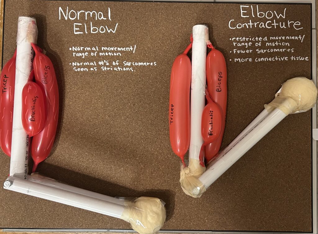
Tyler Benefield’s project focuses on muscle contractures and how they relate to normal muscle contraction. A contracture is when a muscle becomes permanently shortened and stiff, which limits movement. She uses pvc pipes and balloons for her model to show the difference between a normal elbow and one affected by contracture.
Her essay explains how a normal muscle contraction happens. A nerve sends a signal to the muscle, releasing a chemical called ACH. This activates the muscle cell and causes calcium to be released. Calcium allows the proteins actin and myosin to attach and pull on each other, creating the “power stroke” that shortens the muscle. When the signal stops, ACH is broken down, calcium returns to storage, and the muscle relaxes.
She also explains in her essay how contractures form, especially from neurological problems like stroke, cerebral palsy, multiple sclerosis, spinal cord injuries, and traumatic brain injuries. These conditions can cause spasticity, which means the muscles stay tight because of overactive nerve signals. Spasticity does not directly cause contractures, but long-term tightness leads to physical changes in the muscle. Over time, muscles lose some of their normal fibers, become stiff, cannot stretch fully, and develop thickened connective tissue. These changes eventually become permanent, creating a contracture. I thought her model was very creative and helped me understand better.