 Posted by
Logan Kirkland
Steam project essay
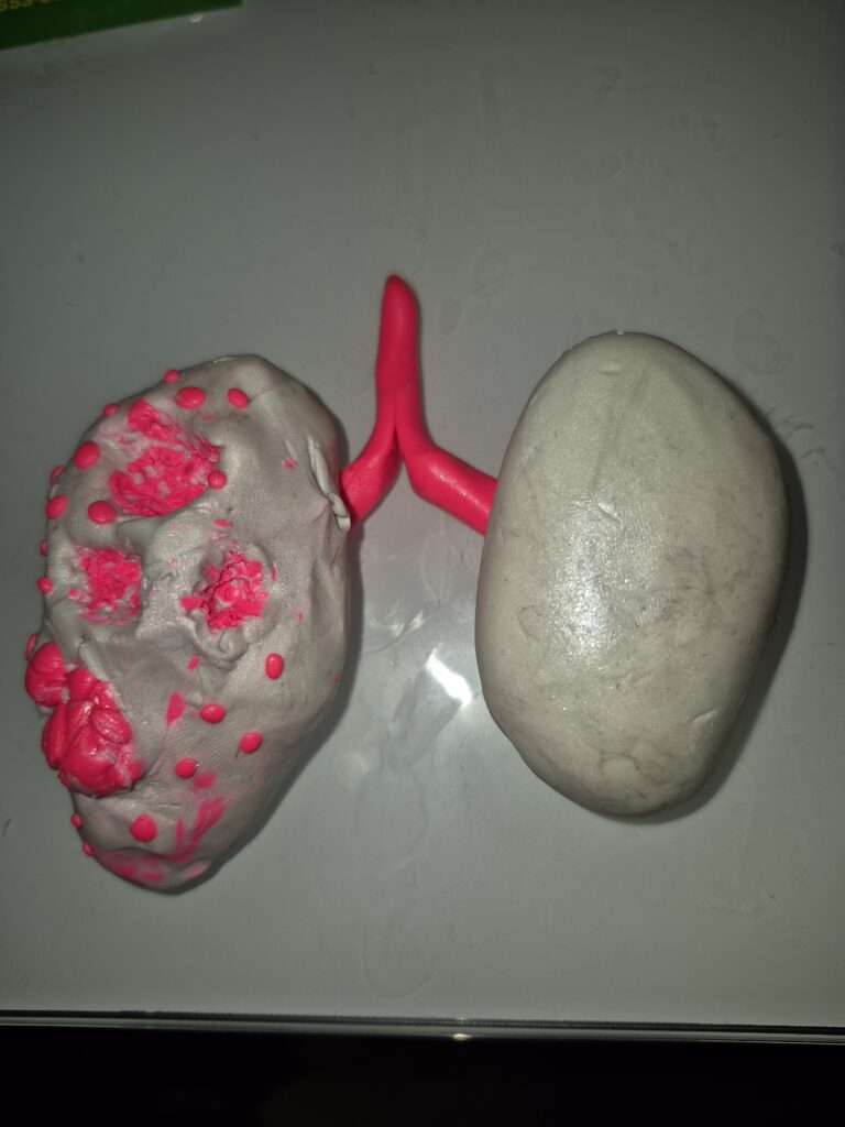
This project will cover the structural and functional effects on the respiratory system caused by the bacterium Mycobacterium tuberculosis. For my project I chose to create a clay model illustration of the difference between a healthy lung and one inflicted with late stage tuberculosis. My model focuses on the physical effects on the lungs and addresses objective 46: “Analyze the effects of disease on function and structure of the respiratory system.” I wanted to make a clay model because it can show the depth of growths and cavities as the infection spreads.
The model demonstrates the differences in structural appearance from a healthy and infected lung. The right lung represents a baseline of healthy respiratory anatomy, shown by a smooth cleaner surface. In contrast the left lung demonstrates the damaging impact of tuberculosis (Ravimohan et al., 2018). I used red clay to show where the infection is attacking healthy tissue and scarring.
The most notable changes are the cavitary lesions. These cavities are caused by the immune response causing caseous necrosis tissue death (Hunter, 2024). This dead tissue is then coughed out leaving holes in the lungs. The small red dots across the surface represent granulomas and military tubercles. These form when the immune system attempts to wall off the bacteria with lymphocytes and macrophages (Alsayed & Gunosewoyo, 2023). These walls replace the soft flexible tissue in the lungs with hard lumps making it even more difficult to breathe. Without a healthy structure, the respiratory system just can’t do its job of keeping the person alive.
The holes and granulomas reduce surface area in the lungs and hinder oxygen exchange. The destruction of lung tissue reduces surface area in the lungs and alveoli where gas exchange occurs meaning blood flowing through the lungs cannot be properly oxygenated (Maiti & Mishra, 2018). Less oxygen flowing and being available to tissues leads to wasting. Wasting is the gradual loss of strength and body mass because the body doesn’t have enough resources to keep the mass (Ravimohan et al., 2018). The shriveled off white texture and redness represents scarring and the lungs loss of compliance; the ability for the lungs to stretch and expand. Fibrous scarring and tissue death makes it significantly harder for the lungs to expand and take in a full breath (Alsayed & Gunosewoyo, 2023).
I represented the bronchial pathways and trachea with the red y shaped clay in between the lungs. In advanced tuberculosis, inflammation and debris from dead tissue can obstruct airflow and trigger coughing which is the primary way the pathogen gets into the air (Hunter, 2024). Tuberculosis spreads primarily through the air whenever an infected person coughs, breathes, or speaks. According to the World Health Organization (2023), TB is the world’s most deadly infectious disease, killing approximately 1.3 to 1.5 million people annually.
In conclusion, this model made of clay acts as a visual representation of objective 46. It demonstrates the delicate structure of the lungs and how a disease can cause catastrophic damage to its function. When looking at the healthy vs diseased side you can see exactly how the disease eats away at the lung tissue. When tuberculosis creates cavities and granulomas it causes catastrophic effects on the lungs’ ability to perform gas exchange.

References
Alsayed, S. S. R., & Gunosewoyo, H. (2023). Tuberculosis: Pathogenesis, current treatment regimens and new drug targets. International Journal of Molecular Sciences, 24(6), 5202. https://doi.org/10.3390/ijms24065202
Hunter, R. L. (2024). Pathology of tuberculosis: How the pathology of human tuberculosis informs and directs animal models. Pathogens, 13(1), 45. https://doi.org/10.3390/pathogens13010045
Maiti, M., & Mishra, A. K. (2018). Structure and function of the tuberculous lung: Considerations for inhaled therapies. Tuberculosis (Edinburgh, Scotland), 91(1), 67–70. https://doi.org/10.1016/j.tube.2010.08.008
Ravimohan, S., Kornfeld, H., Weissman, D., & Bisson, G. P. (2018). Tuberculosis and lung damage: From epidemiology to pathophysiology. European Respiratory Review, 27(147), 170077. https://doi.org/10.1183/16000617.0077-2017
Schoeffel, A., & Panteleimonitis, S. (2020). Portraying tuberculosis through Western art, 1000–2000 CE. Journal of Clinical Medicine, 9(7), 2110. https://doi.org/10.3390/jcm9072110
World Health Organization. (2023, November 7). Tuberculosis. https://www.who.int/news-room/fact-sheets/detail/tuberculosis
The project looks at how Mycobacterium tuberculosis affects the structure and function of the respiratory system. The objective of the project is to show how tuberculosis changes lung structure and explain how these changes affect breathing and gas exchange.
The model compares a healthy lung with a lung affected by late-stage tuberculosis. The healthy lung has a smooth surface and normal structure, while the infected lung shows clear damage, including cavities, granulomas, and scarring. These changes happen because of the immune response to the bacteria. One of the main processes is caseous necrosis, where infected tissue dies and is removed, leaving holes in the lung. Granulomas form when the immune system tries to contain the infection, but they replace flexible lung tissue with harder structures.
These structural changes make it harder for the lungs to function properly. The loss of alveoli and surface area reduces how much oxygen can be exchanged into the blood. Scarring also makes the lungs less compliant, meaning they cannot expand as easily during breathing. This leads to reduced breathing efficiency. In more advanced cases, inflammation and debris can also block airways, which contributes to coughing and helps spread the bacteria.
Model shows that the effects of tuberculosis are not just local damage but a progressive breakdown of how structure supports function in the respiratory system. Once that relationship is disrupted, multiple parts of breathing efficiency fail at the same time.