 Posted by
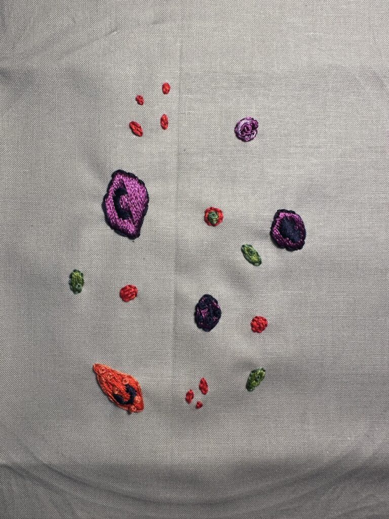
The objectives covered within my STEAM project are to “describe the various WBC and their function,” and “describe disorders of blood and how they can be treated,” from units ten and eleven of this course. This embroidered piece displays several different representations of white blood cells, erythrocytes, platelets, and malaria parasites. This piece is meant to be used as a visual aid when describing the diagnosis of malaria infection using white blood cell counts and blood smears.
Malaria is a parasitic disease transmitted by infected female Anopheles mosquitoes (Cai et al., 2025). According to the World Health Organization (WHO, 2025), there were an estimated 282 million malaria cases worldwide in 2025, 610,000 of which resulted in death. Five species of parasites are known to cause human malaria infection, all under the genus Plasmodium (Mandala et al., 2021). Vaccination and treatment of malaria is a difficult task due to the complicated life cycle of Plasmodium species. After a female takes a blood meal, sporozoites migrate from the mosquito’s salivary gland to the newly infected individual’s blood stream (Mandala et al., 2021). Within two to thirty minutes these sporozoites travel through the blood stream to, “reach and infect liver cells where they multiply to form merozoites” (Mandala et al., 2021, p. 2). As merozoites propagate, this will cause infected liver cells to burst, sending these newly formed parasites back into the bloodstream, where they will now infect red blood cells (Mandala et al., 2021). During the erythrocytic cycle, merozoites inside infected erythrocytes will develop into trophozoites, then finally schizonts (Mandala at al., 2021). It is during this last stage that patients will exhibit the most symptoms of malaria (Mandala et al., 2021). As malaria parasites infect erythrocytes, they can easily travel to all the major organs in the body.
Early diagnosis is very important in containing malaria transmission. Blood smear microscopic examination and rapid diagnostic test strips are the most common tools used for malaria diagnosis at present (Cai et al., 2025). Both processes experience high rates of misdiagnosis, and blood smears depend, “heavily on the experience of laboratory personnel,” and suffer from, “time-consuming processes,” (Cai et al., 2025, p. 3440). Overall, a new process for rapid, cost efficient, and accessible malaria diagnosis is needed. Cai et al. (2025) proposes one way in which professionals could identify malaria infection through the segmentation and classification of white blood cells. According to Mandala et al. (2021), “natural protective immunity against malaria takes years to develop, even in high transmission areas despite repeated exposure to the parasite,” due to the complicated life cycle of the parasite (p. 2). Different antibodies function to target malaria parasites during different stages of the parasite’s life cycle. By determining the presence of different antibodies or white blood cell amounts, one could use that information to diagnose malaria.
Another test for malaria proposed by Burnett et al. (2017) uses haemozoin. Haemozoin (Hz) is a pigment, “generated within infected red blood cells by the malaria parasite,” (Burnett et al., 2017, p. 1). Previously Hz has been used as a malarial biomarker when observing erythrocytes, but the pigment can also be found on white blood cells. It is due to this that red blood cell counts in previous studies may be inflated (Burnett et al., 2017). Using Hz identification within white blood cells could improve misdiagnosis rates.
Malaria is a disease that relies heavily on rapid diagnosis and transmission prevention, which is why new methods of diagnosis are needed. Studies mentioned in this paper offer solutions using the observation of white blood cells, spanning out from the status quo monitoring of erythrocytes. There is much to be improved on in the world of malaria diagnosis and treatment, but there are several different angles to look from. Further studies should strive for speed, accessibility, and cost efficiency.