 Posted by
My objective is to know the structure of the knee joint and compare this joint type to other 5 joint types. Wisdom teeth, popularly known as third molars, are the teeth that are removed by dental practitioners. Many people have them removed because they are the last set of teeth to appear and by the time they do, our jaws are usually too small for their proper growth (Coulthard et al., 2014). In this case, there are surgical techniques used for the removal of lower wisdom teeth when these teeth become impacted. Such impaction can lead to a situation where a tooth is stuck inside the gum and cannot come out properly (Coulthard et al., 2014). They may only partially emerge or grow in at an awkward angle, causing discomfort, swelling, infection, and even damaging other teeth. Similarly, impacted wisdom teeth may lead to less cleaning of the teeth which can cause long-term gum problems (Coulthard et al., 2014). Since our mouth is not big enough to accommodate these teeth either way, an extraction of the tooth becomes both a preventive and corrective measure to problems that have already begun (Coulthard et al., 2014).
The other major reason that people have their wisdom teeth removed deals with proximity to important nerves and bones in the jaw. Research involving imaging for removing lower wisdom teeth indicates that dentists must check how the roots of the tooth align with the mandibular canal, housing a very important nerve (Flygare, L., &; Anders Öhman, 2008). This imaging before surgery helps the dentist avoid nerve damage and also provides them with an idea about the difficulty of the surgery (Flygare, L., & Anders Öhman, 2008). This also helps them understand the anatomy of the shape of the roots or any kinds of twists that may make the procedure more difficult (Flygare, L., & Anders Öhman, 2008). It goes to show that removing a wisdom tooth is not simply a case of pulling it out, but a very delicate procedure that first requires understanding the structure before making any moves on it (Flygare, L., &; Anders Öhman, 2008; Coulthard et al., 2014).
Wisdom teeth removal can greatly impact the overall health of the oral cavity. Once the teeth are removed, the oral cavity has fewer deep gum folds for food and bacteria to be caught. This decreases the risk of infections. The removal of such areas also tends to allow the stability of other teeth around them and decreases the chances of periodontal issues (Coulthard et al., 2014). Discomfort from the surgery healing can occur in many ways, such as swelling or soreness, but the health of the mouth often benefits in the long run. Even radiographic studies confirm that advance planning using proper X-rays results in greater outcomes with less complication, showing again how structure impacts treatment decisions (Flygare, L., &; Anders Öhman, 2008).
To gain a better understanding of all this, it is necessary to consider other parts of the body, such as the joints. The knee joint is considered a synovial hinge joint, which is just a fancy way of saying that it allows the bending and straightening of the leg (Joints in the Human Body: Anatomy, Types &; Function, n.d.). This was designed to be strong and steady with the allowance of mobility for walking and daily activities.
When I compare the knee hinge joint to the five other types of synovial joints, I can really see how different body parts are designed for different purposes. Ball-and-socket joints, such as the shoulder or hip, allow the most movement. Pivot joints help you rotate, such as when you shake your head “no.” Condyloid joints move in two directions, and saddle joints also move in two directions but fit together in a special curved way. Gliding joints let two flat bones slide past each other with small movements (Joints in the Human Body: Anatomy, Types & Function, n.d.). These comparisons make it clear that the structure of a joint determines what it can do, just like how the structure of the jaw and the position of the wisdom teeth determine whether they need to be removed (Flygare, L., & Anders Öhman, 2008; Coulthard et al., 2014).
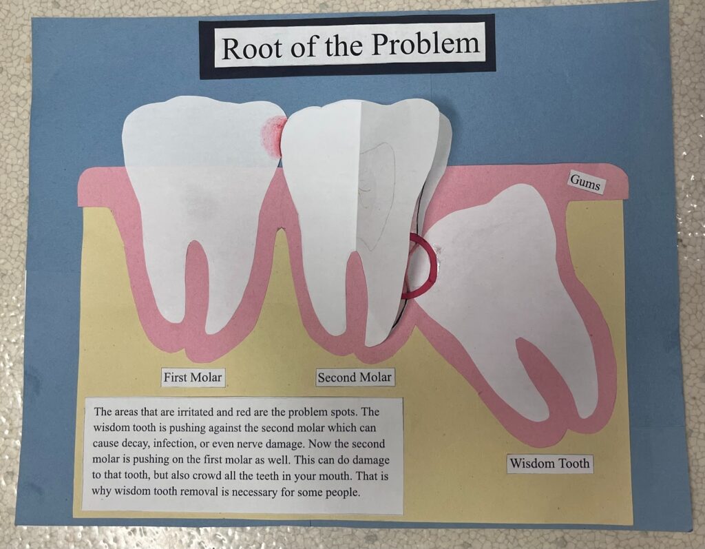
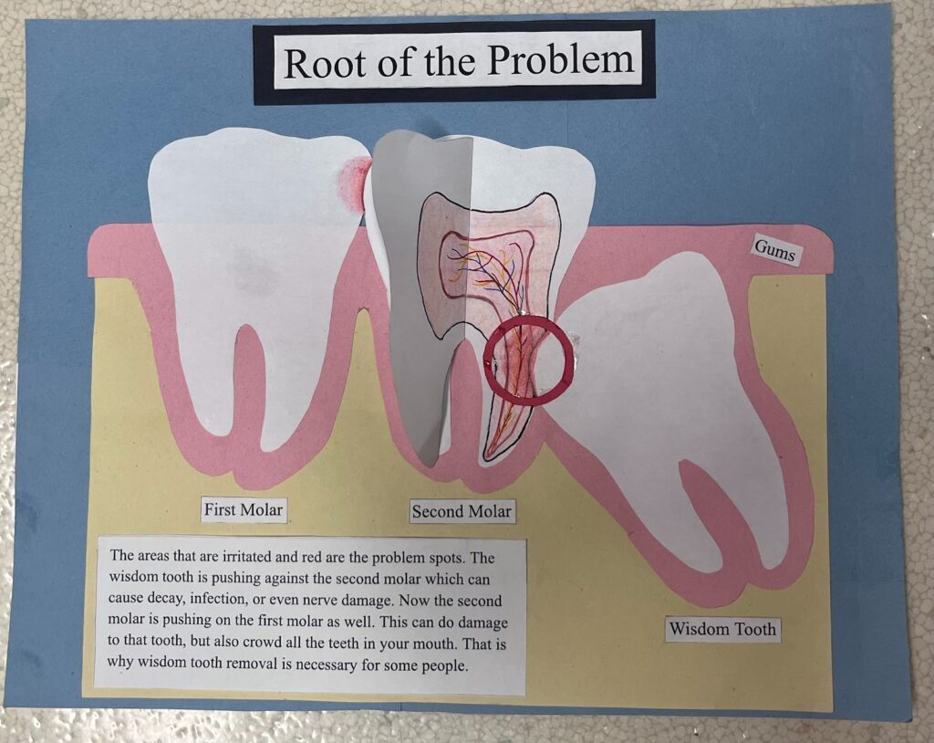
My artwork is a poster that illustrates when the wisdom tooth impacts the second molar and causes problems. These problems could be tooth decay, infection, or nerve damage. The second molar pushes on the first molar making it difficult to keep any spacing between the teeth. When this happens the other teeth in the mouth get crowded, causing cavities and gum disease. In my poster the wisdom tooth is at an angle, but they can range anywhere from being vertical, to completely horizontal. It is important to remove the wisdom teeth for these many reasons.
References
Cleveland Clinic. (2023, July 18). Joints in the Human Body: Anatomy, Types &; Function. Cleveland Clinic. https://my.clevelandclinic.org/health/body/25137-joints
Coulthard, P., Bailey, E., Esposito, M., Furness, S., Renton, T. F., &; Worthington, H. V. (2014). Surgical techniques for the removal of mandibular wisdom teeth. Cochrane Database of Systematic Reviews. https://doi.org/10.1002/14651858.cd004345.pub2
Flygare, L., &; Anders Öhman. (2008). Preoperative imaging procedures for lower wisdom teeth removal. 12(4), 291–302. https://doi.org/10.1007/s00784-008-0200-1

Your explanation really illustrates how the anatomy of the teeth and joints works and why specific procedures within medicine are needed. Most impacted teeth are wisdom teeth, the last molars coming into view, and quite literally, there is a lack of room within the jaw for these teeth. Poor positioning can lead to pain, infection, gum disease, and damage to adjacent teeth. You explain succinctly how important imaging is pre-operatively since dentists need to understand the orientation of the root and proximity to the mandibular canal in order to avoid nerve injury. It allows one to appreciate that a removal of a wisdom tooth is actually a delicate surgical procedure and not merely a tooth extraction.
This directly relates to the knee joint, reinforcing that structure determines function within the body. The knee is a synovial hinge joint, built to be strong and stable yet allows flexion and extension. Compared to other synovial joints-the ball-and-socket, pivot, condyloid, saddle, and gliding joints-one can quickly realize that different joints are shaped for specific movements. Much like the structure of the joints dictates what kind of movement one can have, the position and structure of the wisdom teeth will determine whether or not they will need removal.
Your art ties these ideas together visually by demonstrating how such an impacted tooth can crowd or damage the molars next to it. With your poster, you are able to show different angles of impaction and why early removal can prevent complications such as decay, infection, nerve damage, and gum disease. Overall, your discussion relates anatomy to function, and also the importance of understanding structure in both dental procedures and joint movement.