 Posted by
Different joints are classified by whether they are directly connected to fibrous connective tissue or cartilage or if the articulating surfaces have contact in the fluid filled joint cavity (Betts et al., 2012). The synovial joint is the most common joint, this joint supports movement between adjacent bones. The two bones are connected through the articular capsule which holds the articular cartilage and synovial fluid (Betts et al., 2012). The joint cavity is what defines the structure of the synovial joint. There are several types of joints under the synovial joint umbrella. The knee is a hinge joint, which is a type of synovial joint, where specifically the convex end of one bone articulates with the concave end of the adjoining bone. This hinge joint supports bending and straightening motions on a single axis (Betts et al., 2012). The pivot joint is a rounded portion of a bone enclosed with a ring formed by articulation, the bone rotates within this ring. The pivot joint can be found in the vertebrae and allows for turning the head from side to side. The saddle joint differs from this structure with two articulating surfaces possessing a saddle shape, being concave and convex in opposite directions. An example of the saddle joint is the movement of the thumb away from the palm (Betts et al., 2012). The plane joint also known as the gliding joint is found between the tarsal bones. This joint has articulating surfaces which can have a slight curve or flat and slide easily against each other. Plane joints can support multiple movements depending on where they are in the body, they may only have one type of movement function (Betts et al., 2012). The ball and socket joint is that which forms the hip joint, connecting the head of the femur with the hip bone. This joint is similar to the plane joint in that it is multiaxial, which means it allows movements within three planes (Betts et al., 2012).
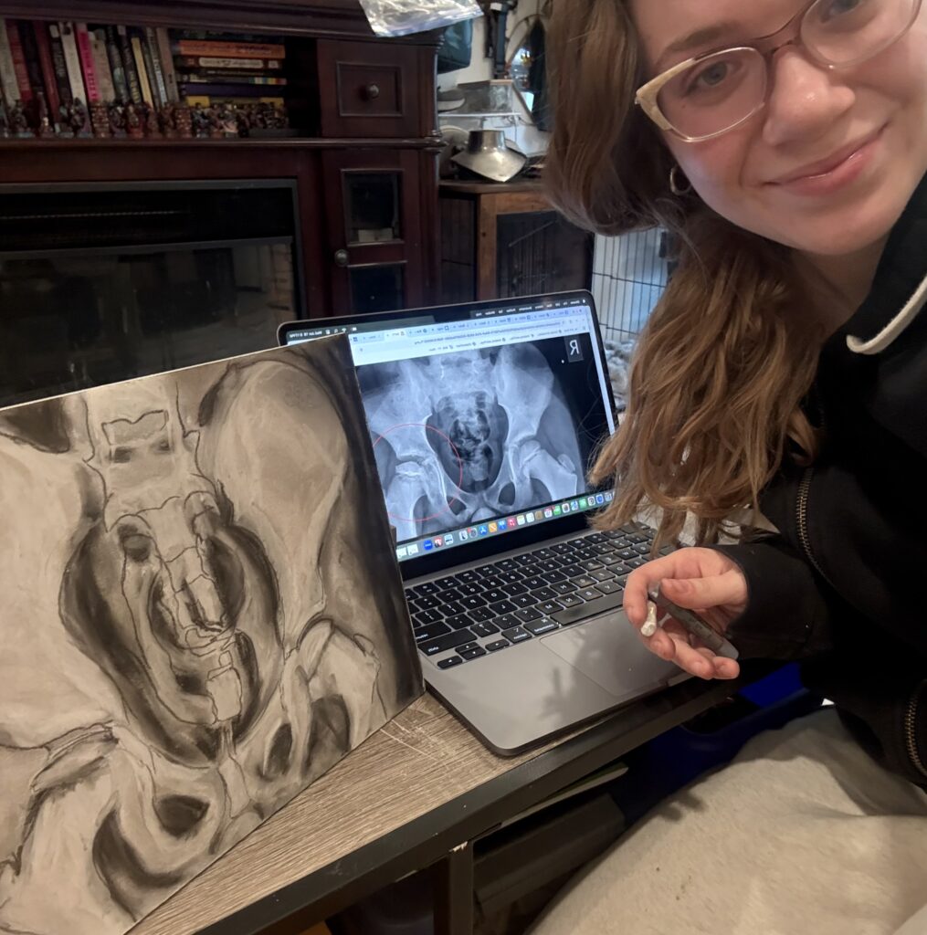
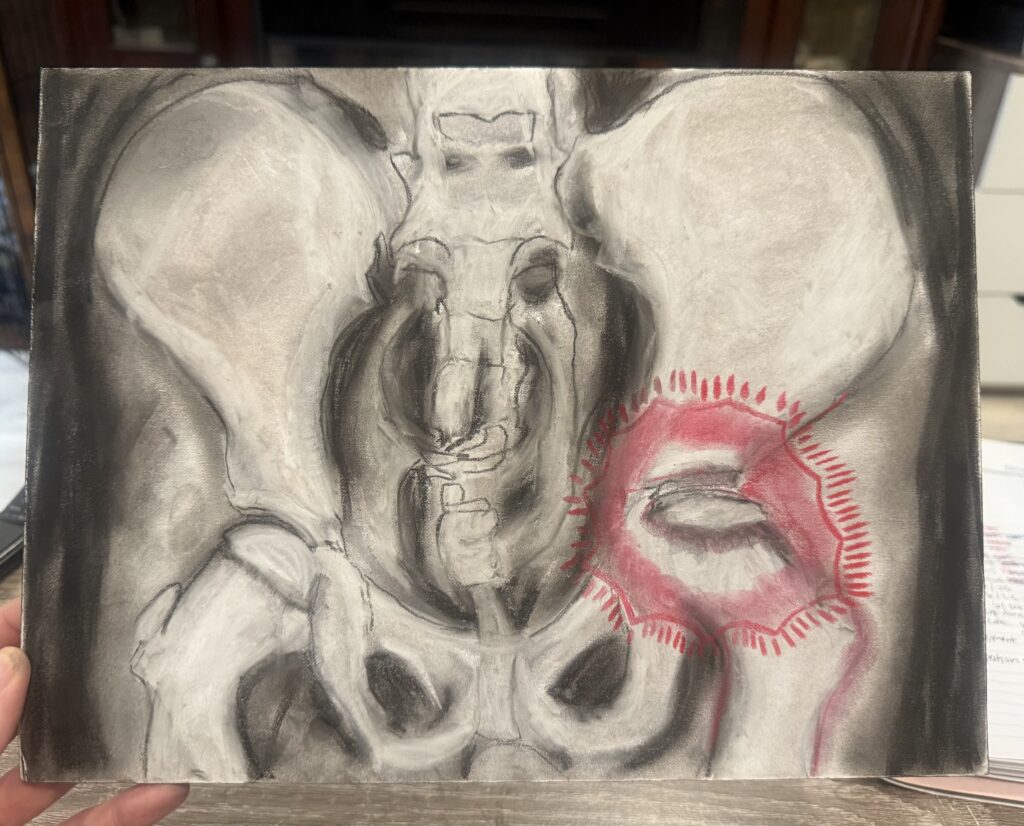
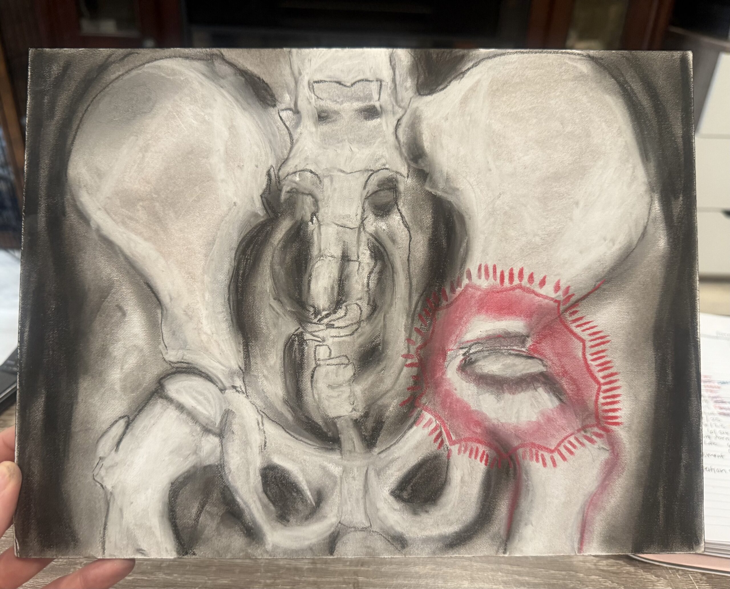
Perthes also known as Legg-Calve Perthes, is a disease that develops in children defined as the unilateral or bilateral necrosis of the femoral head. This femoral head is what is surrounded by the socket portion of the hip joint, the acetabulum. This is what forms the ball and socket joint, both the femur and the hip bone connect through articular cartilage and capsule which is surrounded by several ligaments. The iliofemoral, pubofemoral, and ischiofemoral ligament all thickly cover the head and neck of the femur (Betts et al., 2012). Bone cells, osteoclasts and osteoblasts decay under necrosis which leads to microfractures.Overall the deformities in the femoral head are greater when the force applied to the femoral head is greater than its capacity to resist deformation. With the calcium increase in the dying bone, it is more susceptible to microdamage. When the femoral head loses density the height actually falls, leaving the femoral head significantly shorter and flattened (Rodríguez-Olivas et al., 2022). Due to the force regularly applied to the hip joint, the femoral epiphysis can be irreversibly damaged (Joseph et al., 2023). Perthes can result in irregular function of the ball and joint socket, which presents as a loss of range of motion, lameness, and a limp (Rodríguez-Olivas et al., 2022). Perthes is most frequently found in children between the ages of 2 and 16, with the peak age of onset being between 5 and 6 years old. The interesting piece about perthes is that the exact cause of vascular disruption which leads to femoral epiphyseal necrosis is still unknown (Joseph et al., 2023). It is believed that legg-calve perthes disease is caused by a combination of factors including environmental, metabolic, and genetic, inflammation has also shown to contribute to the development (Rodríguez-Olivas et al., 2022).
References
Betts, J. G., Desaix, P., Johnson, E., Johnson, J. E., Korol, O., Kruse, D., Poe, B., Wise, J. A., Womble, M., Young, K. A. (2012). Anatomy and Physiology. OpenStax Rice University (pp. 314, 320-326) https://openstax.org/books/anatomy-and-physiology/pages/1-introduction
Joseph, B., Shah, H., & Perry, D. C. (2023). Epidemiology, natural evolution, pathogenesis, clinical spectrum, and management of Legg–Calvé–Perthes. Journal of Children’s Orthopaedics (British Editorial Society of Bone & Joint Surgery), 17(5), 385–403. https://doi.org/10.1177/18632521231203009
Rodríguez-Olivas, A. O., Hernández-Zamora, E., & Reyes-Maldonado, E. (2022). Legg–Calvé–Perthes disease overview. Orphanet Journal of Rare Diseases, 17(1). https://doi.org/10.1186/s13023-022-02275-z