
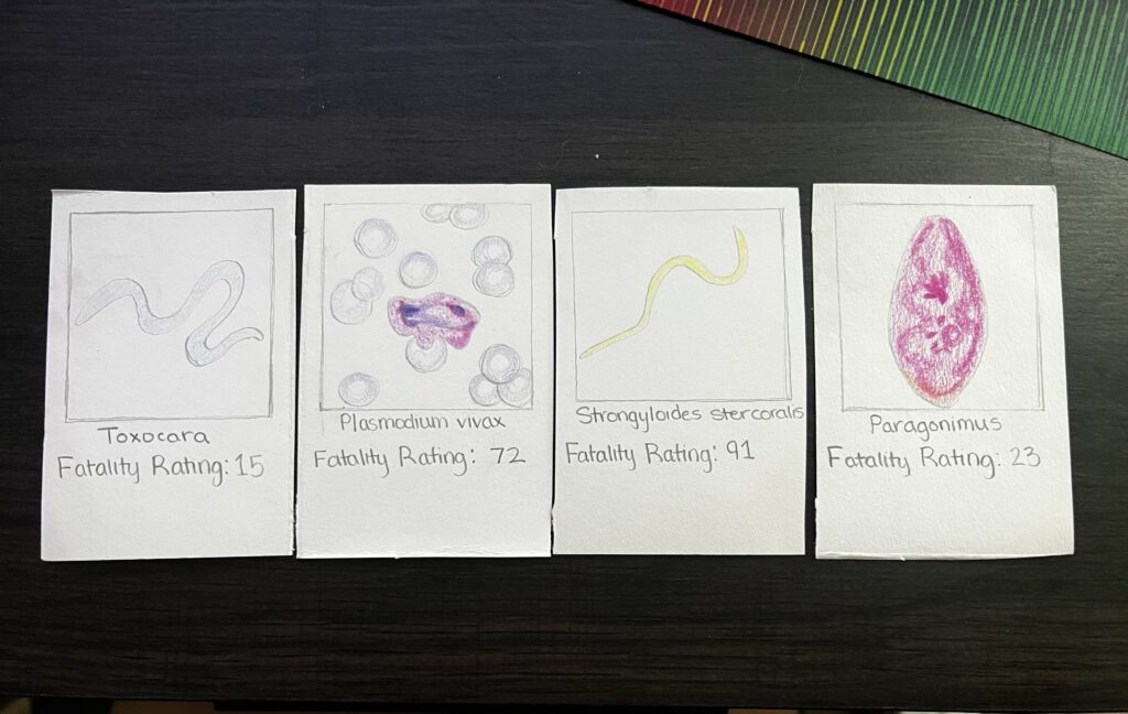
Malaria
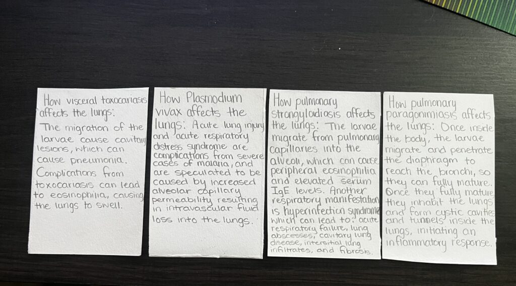
Malaria, which is most commonly spread by mosquitoes in underdeveloped countries, can be a fatal parasitic disease. After being bit by a mosquito carrying malaria, the parasites then travel to your liver where they lie dormant. The parasites usually remain dormant for about ten days, but can remain dormant for up to two weeks. After this period of dormancy, the parasites leave the liver and infect red blood cells, which causes symptoms to appear. Acute lung injury (ACL) and acute respiratory distress syndrome (ARDS) can be symptoms of malaria. ARDS results from severe complications from Malaria, and most commonly affects pregnant women and non-immune individuals.Increased alveolar capillary permeability resulting in intravascular fluid loss into the lungs appears to be the key pathophysiologic mechanism (Mohan et al., 2008). Pneumonitis may also contribute to respiratory distress.
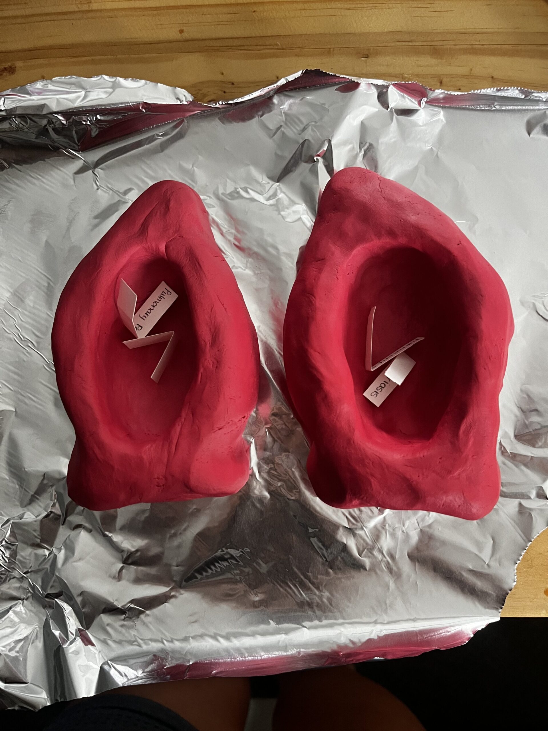
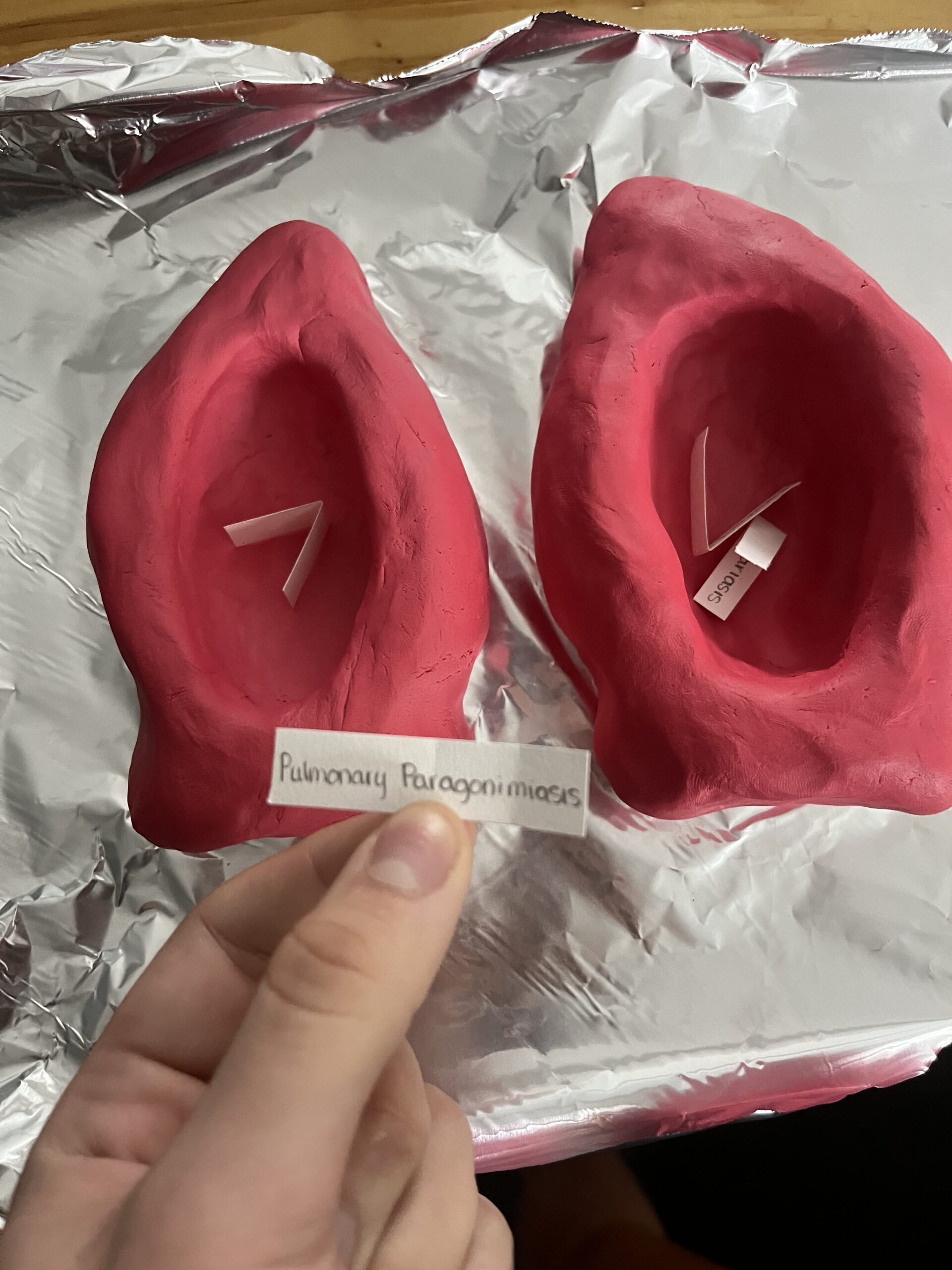
Pulmonary Paragonimiasis
Paragonimus is a lung fluke that affects the lungs after ingesting undercooked seafood. Once inside the body, metacercariae excyst in the small intestine and release larvae. These larvae penetrate the duodenal wall and enter the peritoneal cavity. The larvae then go through a migration period, which usually takes about a week, where they will then penetrate the diaphragm, enter the pleural cavity, and then migrate through lung tissue to enter the bronchi. The maturation process of these larvae take about 5 to 6 weeks, and develop into adults inside of cystic cavities that they formed inside the bronchi. Once the larvae fully mature, they inhabit the lungs, and by doing so they form cystic cavities and tunnels inside the lungs, which initiates an inflammatory response. Symptoms of pulmonary paragonimiasis resemble chronic bronchitis or tuberculosis with blood-tinged sputum containing the eggs of the parasite, chest pain and/or shortness of breath, diarrhea, and abdominal pain. People infected with pulmonary paragonimiasis have very mild symptoms, and if left untreated, people can live with the infection for decades, making it not so fatal.
Pulmonary Strongyloidiasis
Pulmonary strongyloidiasis is a parasitic infection caused by coming into contact with infective filariform larvae on the skin. These larvae are found in contaminated soil. After coming into contact with the larvae, they penetrate the skin, where they then travel through the circulatory system into the lungs and alveoli. They continue to travel to the upper airway until they are swallowed, and end up in the small intestine where they will stay to mature. The adult female parasite then starts laying eggs, most of which will be excreted through stool. The migration from pulmonary capillaries into the alveoli can cause peripheral eosinophilia and elevated serum IgE levels. Another respiratory manifestation associated with pulmonary strongyloidiasis is hyperinfection syndrome, this phase of infection commonly leads to acute respiratory failure, lung abscesses, cavitary lung disease, interstitial lung infiltrates, and fibrosis (Mokhlesi et al., 2010).
Visceral Toxocariasis
Toxocariasis is a parasitic infection caused by roundworms, and is spread by animal feces in contaminated soil. After the eggs are ingested they travel from your intestines to the liver or the lungs. Symptoms of visceral toxocariasis include: fever, cough, wheezing, fatigue, abdominal pain, skin rash, enlarged liver or spleen, and pneumonia. People infected with toxocariasis usually experience no symptoms or very mild symptoms, except for in severe cases where pneumonia develops. The severity of symptoms depends heavily on the cavitary lesions caused by the migration of the larvae. Complications from toxocariasis can lead to eosinophilia, which causes the lungs to swell.
Izzy’s Project was on a few parasitic infections that have an effect on the lungs. She first talks about Malaria which she describes as a potentially fatal disease spread by mosquitos. After being bitten by a mosquito, the parasite travels to the liver where it remains dormant for up to two weeks then it infects red blood cells. This causes Acute lung injury and acute respiratory distress syndrome. The next parasitic infection she talks about is Pulmonary Paragonimiasis, a lung fluke that affects the lungs after eating raw or undercooked seafood. She then goes into the path the flatworm takes. This parasite enters the body as metacercariae which releases larvae into the small intestines. The larvae then penetrate the duodenal wall and travel to the peritoneal cavity and migrate through the diaphragm to the pleural cavity, where they eventually reach the lungs. Over the course of 5-6 weeks, they mature into adults with cystic cavities in the bronchi causing an inflammatory response. Symptoms she listed include chronic bronchitis-like symptoms, blood-tinged sputum with parasite eggs, chest pain, shortness of breath, diarrhea, and abdominal pain. The next parasitic infection she talks about is Pulmonary Strongyloidiasis, which is caused by contact with larvae in contaminated soil. These larvae then penetrate the skin to travel to the lungs and alveoli via the circulatory system, they eventually reach the small intestines where they mature into adults and lay eggs. Migration from pulmonary capillaries to the alveoli can lead to elevated levels of eosinophils and IgE. This infection can manifest as hyper-infection syndrome, resulting in severe respiratory complications like acute respiratory failure, lung abscesses, and fibrosis. The last parasitic infection she covered was Visceral Toxocariasis, caused by roundworm and spread through contaminated soil and animal feces. The process of infection begins with the ingestion of eggs that migrate to the liver or lungs. Symptoms of this infection include fever, cough, wheezing, fatigue, abdominal pain, skin rash, enlarged spleen/liver, and pneumonia in severe cases. Most infected individuals experience mild to no symptoms but complications can arise due to cavitary lesions caused by larval migration, leading to eosinophilia and lung swelling.